
3D Scan for Root Canal: Finding Hidden Canals
3D Scan for Root Canal: Finding Hidden Canals
“My dentist says I need a 3D scan for root canal. I already had x-rays—isn’t that enough? This seems like an unnecessary expense.”
This frustration is understandable. You’re already facing root canal treatment (which nobody wants), and now you’re being told you need additional imaging.
But here’s what many patients don’t realize: The #1 reason root canals fail is missed canals. Teeth often have more root canals than dentists expect—and standard x-rays simply cannot show these hidden canals because they collapse three-dimensional internal anatomy into a flat, two-dimensional image.
The statistics are eye-opening: Studies show that CBCT 3D scans identify additional canals in 30-40% of teeth that appeared to have “normal” anatomy on standard x-rays. That means nearly 4 out of 10 teeth undergoing root canal treatment have hidden canals that 2D imaging misses entirely.
When a canal is missed during root canal treatment, bacteria remain inside that untreated canal. The tooth continues to be infected. Pain persists or returns. The root canal “fails”—not because the dentist did poor work on the canals they treated, but because they never knew other canals existed.
This comprehensive guide explains why root canal anatomy is more complex than most people realize, what standard x-rays can and cannot show about internal tooth structure, how CBCT 3D scans reveal the complete canal system including hidden branches, which teeth most commonly have unexpected anatomy, and how this imaging technology transforms root canal success rates.
Whether you’re preparing for your first root canal or dealing with a failed previous treatment, understanding why 3D imaging matters helps you appreciate that this isn’t an unnecessary expense—it’s essential insurance against root canal failure.
Understanding Root Canal Anatomy: More Complex Than You Think
Before exploring imaging, let’s understand why finding all root canals is so challenging.
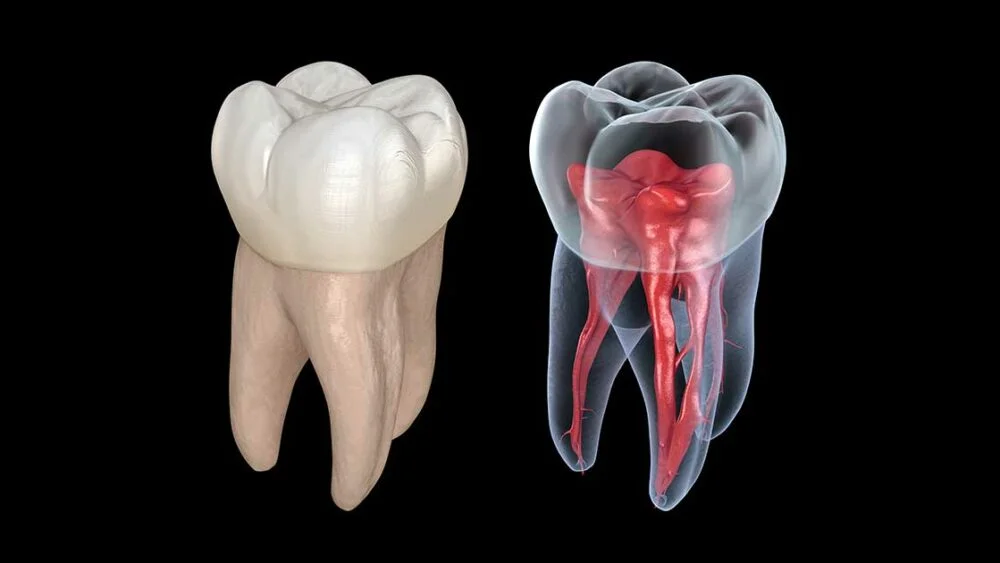
What Root Canals Actually Are
Root canals are tiny passageways inside your tooth roots that contain the pulp—nerves, blood vessels, and connective tissue keeping the tooth alive.
When pulp becomes infected (from deep decay, cracks, or trauma), it must be removed. Root canal treatment involves:
- Accessing the pulp chamber inside the tooth
- Finding and entering all root canals
- Removing infected pulp tissue
- Cleaning and shaping canals
- Filling canals with sealing material
- Restoring the tooth with a crown
The critical step is “finding and entering all root canals.” If even one canal is missed, bacteria survive in that untreated space, and infection persists.
Why Canal Anatomy Varies So Much
Dental textbooks show “typical” root canal anatomy for each tooth type. For instance, upper first molars “typically” have 3 roots and 4 canals. Lower molars “typically” have 2 roots and 3 canals.
The problem? Many teeth don’t follow the textbook.
Anatomical variations include:
- Extra canals: A tooth expected to have 3 canals might have 4, 5, or even 6
- Branching canals: A single canal might split into two separate branches
- Lateral canals: Tiny canals branching off the main canal sideways
- Accessory canals: Small additional canals connecting to the main system
- C-shaped canals: Instead of separate round canals, one large C-shaped canal
- Isthmuses: Web-like connections between canals that also harbor bacteria
These variations occur in predictable patterns based on tooth type and patient ethnicity, but they’re invisible on standard x-rays—making them “hidden” from dentists working with 2D imaging.
The Consequences of Missed Canals
“What actually happens if a canal is missed during my root canal?”
The missed canal remains infected. Bacteria living in that untreated space continue causing problems:
Persistent symptoms: Pain, sensitivity, or swelling that never fully resolve after root canal treatment.
Chronic infection: Low-grade infection persists, potentially causing bone loss around the root tip.
Treatment failure: Eventually, the tooth becomes symptomatic again—sometimes months or years later.
Need for retreatment: The tooth requires root canal retreatment where the dentist must find and treat the missed canal—more complex and expensive than getting it right initially.
Possible extraction: If retreatment fails or isn’t feasible, the tooth may require extraction and replacement.
Studies show missed canals cause 30-50% of root canal failures. This isn’t about dentist skill in treating canals they found—it’s about finding all canals in the first place.
For expert root canal treatment in Gandhinagar, comprehensive CBCT imaging ensures complete canal identification before treatment begins.
💡 Quick Tip: If your dentist finds a tooth needs root canal treatment and mentions the tooth might have “complex anatomy” or “extra canals,” this is exactly when CBCT imaging provides critical value. Don’t view it as an unnecessary expense—view it as insurance against treatment failure.
🔑 Key Takeaway: Root canal success depends on finding and treating ALL canals. Standard x-rays show some canals but miss many variations. CBCT 3D imaging reveals the complete internal anatomy, dramatically reducing the risk of missed canals.
What Standard X-rays Show About Root Canals (and What They Miss)
Understanding the limitations of 2D imaging helps you appreciate why CBCT makes such a difference for root canal treatment.
Standard Dental X-rays for Root Canals
Periapical x-rays (the small x-rays where you bite on a sensor) are the standard imaging for root canal evaluation.
What these x-rays show well:
- Number of roots the tooth has
- Approximate root length
- Obvious root canal infections (dark areas at root tips)
- General root anatomy
- Some canal curvatures
What they show poorly or not at all:
- Exact number of canals within each root
- Canals positioned directly behind or in front of each other
- Very thin or branching canals
- C-shaped canal configurations
- Isthmuses connecting canals
- Three-dimensional root curvatures
The Fundamental 2D Limitation
Standard x-rays collapse three-dimensional anatomy into a two-dimensional image. Imagine trying to understand the interior layout of a building from a single photograph of its exterior—you see the outside but have no idea about the internal room configuration.
Specific limitations for root canals:
Superimposition: When two canals lie directly in front of or behind each other (from the x-ray beam’s perspective), they appear as one canal on the image. You see one canal on the x-ray, but two exist in reality.
No depth information: X-rays don’t show whether a canal is positioned toward the cheek side or tongue side of the root—critical information for accessing that canal.
Thin canals invisible: Very thin canals or canals with small openings might not appear on x-rays at all, rendering them completely invisible.
No cross-sectional view: You can’t see the shape of the canal in cross-section—is it round, oval, ribbon-shaped, or C-shaped? This affects treatment approach.
How CBCT 3D Scans Reveal Hidden Root Canals
CBCT (Cone Beam Computed Tomography) scanning creates complete three-dimensional reconstructions of tooth anatomy by capturing hundreds of x-ray images from different angles.
What CBCT Shows That Standard X-rays Cannot
Complete canal system mapping: Every canal, branch, and connection becomes visible in three dimensions.
Exact canal positions: CBCT shows whether canals are positioned toward the cheek, tongue, or middle of the root—guiding access during treatment.
Cross-sectional views: You can “slice” through the tooth at any level and see the canal shape in cross-section—revealing C-shaped canals, isthmuses, or unusual configurations.
Canal branching patterns: Secondary canals, lateral canals, and accessory canals appear clearly in 3D imaging.
Number of canals with certainty: CBCT definitively shows whether a tooth has 2, 3, 4, or more canals—no guessing based on 2D projections.
Root curvatures in all dimensions: Three-dimensional curvature is fully visible, not just the curvature visible in one plane on standard x-rays.
Real-World Example: Lower First Molar
Let’s make this concrete with a common scenario.
A lower first molar “typically” has 2 roots and 3 canals (one canal in the front root, two canals in the back root).
On standard x-rays: You see 2 roots and what appears to be 3 canals. The dentist plans standard root canal treatment expecting to find and treat 3 canals.
On CBCT 3D scan: The scan reveals:
- The back root actually contains 3 canals, not 2
- The additional canal (called the “middle mesial” canal) runs between the two expected canals
- This extra canal is positioned directly between the other two, making it invisible on 2D x-rays where it’s superimposed behind one of the visible canals
With CBCT knowledge, the dentist knows to look for that third canal in the back root and successfully treats all canals. Without CBCT, the middle mesial canal would likely be missed—a hidden source of persistent infection.
Studies show 30-60% of lower molars have this middle mesial canal that standard x-rays rarely reveal. CBCT identifies it in virtually every case where it exists.
At our CBCT imaging facility in Gandhinagar, advanced 3D scans provide the complete endodontic roadmap that successful root canal treatment requires.
🔑 Key Takeaway: CBCT doesn’t just show canals more clearly—it reveals canals that are completely invisible on standard x-rays. This discovery rate of 30-40% additional canals transforms root canal success rates from “usually works” to “almost always works.”
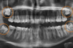
Which Teeth Most Commonly Have Hidden Canals
Certain teeth are notorious for anatomical variations that standard x-rays miss. Knowing which teeth benefit most from CBCT helps you understand why your dentist might recommend 3D imaging.
Upper First Molars: The MB2 Canal
Upper first molars are famous for having an extra canal that’s missed in 50-70% of cases when dentists work without CBCT.
Expected anatomy: 3 roots, 4 canals (one canal in each of two roots, two canals in the third root).
Hidden reality: The root expected to have two canals actually has a fourth canal (called “MB2” – second mesiobuccal canal) in 70-95% of teeth, depending on patient age and ethnicity.
Why it’s missed: The MB2 canal opening is often hidden under a shelf of tooth structure. Without knowing exactly where to look (which CBCT reveals), dentists miss it frequently.
CBCT benefit: Shows precise MB2 location, allowing dentists to remove the overlying tooth structure and access the hidden canal.
Lower Molars: The Middle Mesial Canal
As mentioned earlier, lower molars frequently have an extra canal between the two expected canals in the front root.
This middle mesial canal is often extremely thin and positioned between the other canals—invisible on standard x-rays but clearly visible on CBCT cross-sections.
CBCT benefit: Reveals whether this canal exists and shows exactly where it’s positioned relative to the other canals.
Lower Premolars: Multiple Canal Systems
Lower premolars appear simple on x-rays (one root, one canal) but frequently have far more complex anatomy.
Hidden reality: 20-40% have two separate canals, and some have three canals—all within a single thin root.
Why it’s missed: The canals split from a single chamber into separate branches that may rejoin or remain separate—impossible to determine from 2D imaging.
CBCT benefit: Shows exactly how many canals exist and whether they remain separate or merge, guiding treatment approach.
Upper Premolars: Root Division Variations
Upper premolars sometimes have one root, sometimes two roots, and canal numbers vary accordingly.
Standard x-rays struggle to determine whether one or two roots exist when roots overlap from the x-ray beam’s perspective.
CBCT benefit: Definitively shows root count, canal count within each root, and their three-dimensional configuration.
💡 Quick Tip: If you’re getting root canal treatment on an upper first molar, lower molar, or any premolar and your dentist hasn’t mentioned CBCT imaging, ask whether these teeth commonly have hidden canals. The answer should prompt discussion about whether 3D imaging would benefit your case.
When CBCT Is Most Valuable for Root Canals
Not every root canal requires CBCT imaging. Understanding when 3D imaging provides genuine value helps you evaluate recommendations appropriately.
High-Value CBCT Situations
Teeth known for complex anatomy: Upper first molars, lower molars, all premolars—teeth where hidden canals are common.
Retreatment cases: When a previous root canal failed, CBCT often reveals missed canals that caused the failure.
Persistent symptoms after treatment: If pain continues after root canal therapy, CBCT can identify untreated canals or anatomy explaining the symptoms.
Difficult access cases: When clinical examination suggests canals will be hard to find or access, CBCT provides a roadmap.
Patients with unusual anatomy: Some people have anatomical variations throughout their teeth—if one tooth showed complex anatomy, others likely will too.
Teeth with calcified canals: When canals become partially blocked with calcium deposits, CBCT shows where the canal continues beyond the blockage.
Lower-Value Situations
Simple anterior teeth: Front teeth typically have very straightforward one-canal anatomy that standard x-rays show adequately.
Young patients with developing teeth: Root anatomy is often simpler in younger patients.
Emergency pain relief: When the goal is immediate pain relief by opening the tooth and draining infection, detailed anatomy mapping can wait until definitive treatment.
The decision should be individualized based on tooth type, clinical findings, and treatment goals.
How CBCT Changes Root Canal Treatment
CBCT doesn’t just provide information—it changes what endodontists actually do during treatment.
Treatment Planning Changes
Access opening design: CBCT shows exact canal locations, allowing the dentist to design the access opening that provides direct pathways to all canals—including hidden ones.
Instrumentation approach: Knowing canal curvatures in three dimensions helps select appropriate instruments and techniques.
Cleaning strategy: Complex anatomy like isthmuses or C-shaped canals requires different cleaning approaches than simple round canals—CBCT reveals which approach is needed.
Filling technique: Canal shape and branching pattern influence what filling materials and techniques will seal the system effectively.
Improved Success Rates
Research consistently shows better root canal outcomes when CBCT guides treatment:
Higher success rates: Studies show 5-15% improvement in long-term success when CBCT identifies all canals versus treatment with 2D imaging alone.
Reduced retreatment need: Treating all canals the first time prevents the need for retreatment when hidden canals cause persistent infection.
Fewer extractions: Teeth that might have been extracted as “hopeless” after failed root canal can often be saved when CBCT reveals what was missed initially.
Better patient experience: Avoiding retreatment means less time in the dental chair, lower costs, and better outcomes.
For comprehensive dental care in Gandhinagar, CBCT-guided endodontics represents the modern standard for predictable root canal success.
Comparing Root Canal Approaches: 2D vs. 3D Imaging
| Factor | Standard X-rays Only | CBCT 3D Scan |
|---|---|---|
| Canal Detection | Miss 30-40% of extra/hidden canals | Identify virtually all canals including variations |
| Canal Location | Approximate—guessing involved | Exact position in three dimensions |
| Complex Anatomy | C-shaped canals, isthmuses often unrecognized | All variations visible and planned for |
| Treatment Planning | Based on textbook expectations | Based on actual patient anatomy |
| Access Opening | May need modification during treatment | Designed optimally before treatment starts |
| Success Rate | Good but limited by missed canals | Excellent—all canals treated |
| Retreatment Risk | Higher—missed canals cause failures | Lower—complete treatment first time |
| Patient Confidence | “I hope we got everything” | “We know we treated all canals” |
| Cost | Lower imaging cost, higher failure risk | Higher imaging cost, lower failure risk |
| Professional Standard | Adequate for simple cases | Optimal for complex cases |
CBCT for Root Canal Retreatment
When a previous root canal fails, CBCT is often essential for understanding why and planning successful retreatment.
Common Retreatment Scenarios
Persistent infection despite treatment: Pain continues or returns months/years after root canal.
The CBCT investigation: Often reveals an untreated canal that was missed during the original treatment. Bacteria survived in that hidden canal, causing persistent infection.
Inadequate cleaning of known canals: Sometimes CBCT shows the dentist found all canals but didn’t clean them to the root tip—leaving infected tissue in the bottom portion.
Missed anatomy like isthmuses: CBCT might reveal connections between canals (isthmuses) that weren’t cleaned during original treatment.
Root fractures: CBCT can identify vertical root fractures—cracks extending through the root—that cause treatment failure and usually require extraction.
Retreatment Planning
CBCT allows strategic retreatment planning:
- Identify which canal(s) need retreatment
- Locate the missed canal precisely
- Assess whether retreatment is feasible or extraction is necessary
- Plan surgical approaches if conventional retreatment won’t access the problem area
Without CBCT, retreatment often involves re-treating all canals hoping to stumble upon whatever was missed—less efficient and less predictable than targeted treatment guided by 3D imaging.
💡 Quick Tip: If your root canal failed and your dentist recommends retreatment, ask whether CBCT will be used to identify what was missed. Retreating without understanding why the first treatment failed often leads to second failures.
What to Expect: CBCT-Guided Root Canal Process
Step 1: Initial Evaluation
Dentist diagnoses the need for root canal treatment through examination and standard x-rays. If tooth type or clinical findings suggest complex anatomy, CBCT is recommended.
Step 2: CBCT 3D Scanning
Scan takes 10-20 seconds. Processing creates 3D reconstruction showing complete internal tooth anatomy.
Step 3: Treatment Planning
Dentist analyzes CBCT data:
- Counting canals in each root
- Mapping canal positions in three dimensions
- Identifying branching patterns or unusual configurations
- Planning optimal access opening design
Step 4: Root Canal Treatment
During treatment, dentist follows the anatomical roadmap from CBCT:
- Access opening designed to reach all identified canals
- Each canal is located and entered
- Cleaning and shaping proceeds with knowledge of canal curvatures
- Filling technique selected based on canal configuration
The entire process is more efficient and predictable because the dentist knows exactly what anatomy to expect rather than discovering it during treatment.
Step 5: Post-Treatment Verification
Follow-up x-rays confirm all identified canals were treated and filled. CBCT might be used again if healing doesn’t progress as expected.
At our facility, advanced dental technology including CBCT ensures root canal treatment addresses all canals for optimal success.
Why Nova Dental Hospital for CBCT-Guided Root Canal Treatment
At Nova Dental Hospital, Gandhinagar, root canal treatment benefits from advanced diagnostic imaging when anatomy demands it.
Our approach includes:
In-house CBCT 3D scanning providing complete endodontic roadmap without external referrals.
Expert endodontic analysis by Dr. Happy Patel with experience interpreting CBCT for complex root canal cases.
Comprehensive treatment capabilities from straightforward root canals to complex retreatment requiring CBCT guidance.
Same-day imaging and treatment when possible—consultation, CBCT, and root canal completion in one visit for appropriate cases.
Transparent communication showing you the 3D images and explaining exactly what anatomy your tooth has and why specific approaches are recommended.
Painless treatment focus using modern techniques and anesthesia protocols that make root canal therapy comfortable.
Conveniently located near PDPU and Gift City, serving patients throughout Gandhinagar who expect endodontic care supported by advanced diagnostics.
Visit our Google Business profile to see patient experiences with our root canal treatment.
Frequently Asked Questions
Do all root canals need CBCT scans?
No—CBCT is most valuable for teeth known to have complex anatomy or when treatment complexity warrants it. Simple front teeth with straightforward one-canal anatomy typically don’t require CBCT. However, upper first molars (notorious for hidden MB2 canals), all molars with multiple roots, premolars (frequently have unexpected extra canals), retreatment cases where previous root canal failed, and teeth with persistent symptoms despite treatment all benefit significantly from CBCT imaging. The decision should be individualized. When your dentist recommends CBCT for root canal treatment, it’s because your specific tooth has high probability of hidden canals that 2D imaging cannot detect. Studies show CBCT identifies additional canals in 30-40% of teeth—meaning nearly 4 out of 10 patients have hidden anatomy that could cause treatment failure if not identified and treated.
How much does a 3D scan cost for root canal treatment?
CBCT scans typically cost ₹3,000-6,000 in Gandhinagar. While this adds to root canal treatment costs, consider it in context: root canal retreatment (needed when the first treatment fails) typically costs ₹10,000-20,000 beyond the original treatment cost. If CBCT prevents retreatment by finding all canals initially, it saves money long-term. Additionally, tooth extraction and replacement (needed when root canal ultimately fails) costs ₹25,000-75,000 depending on replacement method. The few thousand rupees for CBCT imaging that prevents these scenarios represents sound investment. Many dental insurance plans cover CBCT when medically necessary for endodontic treatment. Even without insurance coverage, preventing root canal failure through complete canal identification provides value far exceeding the imaging cost.
Can hidden canals cause root canal treatment to fail?
Absolutely yes—missed canals are the leading cause of root canal failure. Studies show 30-50% of failed root canals result from untreated canals that were never found during the original treatment. When even one canal remains untreated, bacteria surviving in that space continue causing infection. The tooth may feel better temporarily after root canal (because the treated canals are cleaned), but infection persists in the missed canal and eventually causes symptoms to return—pain, swelling, or persistent sensitivity. This isn’t about poor treatment quality on the canals the dentist found and treated. It’s about not knowing additional canals existed. CBCT dramatically reduces this failure mode by revealing all canals before treatment begins, ensuring complete bacteria removal from the entire canal system. When all canals are treated, root canal success rates approach 95%, compared to 75-85% when hidden canals are missed.
What’s the difference between CBCT for root canals versus CBCT for implants?
While both use the same CBCT technology, they’re analyzing different anatomical features. CBCT for implants focuses on: bone dimensions (width, height, density), nerve locations relative to implant position, sinus proximity in upper jaw, and optimal implant positioning for final crown. CBCT for root canals focuses on: internal tooth anatomy (canal count and location), canal branching patterns, root curvatures in three dimensions, and identifying calcifications or anatomical variations inside roots. The scan itself captures the same 3D data, but what the dentist analyzes differs based on treatment goals. Both applications dramatically improve treatment success by providing three-dimensional information that 2D x-rays cannot show. Your dentist might use CBCT at different times for different purposes—before implant placement to plan surgery, before root canal to map anatomy, or for other diagnostic needs where 3D visualization provides critical information.
If my dentist doesn’t have CBCT, should I go somewhere else for root canal treatment?
Not necessarily, but it depends on tooth complexity and your dentist’s experience. Simple front teeth with straightforward anatomy don’t require CBCT for successful treatment. However, if you need root canal on upper first molar (notorious for missed MB2 canals), any lower molar (frequently have hidden middle mesial canals), any premolar (often have unexpected extra canals), or any tooth where previous root canal failed, treatment without CBCT guidance carries higher failure risk. In these situations, consider seeking treatment from an endodontist (root canal specialist) with CBCT capabilities, or ask your general dentist whether they can refer you for CBCT imaging before performing the root canal. Many general dentists successfully treat complex root canals, but access to CBCT when anatomy demands it significantly improves outcomes. The key question is whether your specific tooth has high probability of hidden anatomy—if yes, CBCT-guided treatment is worth seeking.
Related posts


Write a Comment
Recent Posts

3D Scan for Root Canal: Finding Hidden Canals

Making Wisdom Tooth Removal Safer with CBCT Scans

X-rays Before Dental Implants: Using 3D Tech for Success

