
Making Wisdom Tooth Removal Safer with CBCT Scans
Making Wisdom Tooth Removal Safer with CBCT Scans
“My dentist says my wisdom tooth is close to a nerve and I need a 3D scan before extraction. Isn’t that overkill?”
This question reflects a dangerous misconception—that careful surgical technique alone can compensate for incomplete diagnostic information.
Here’s what many patients don’t realize: The difference between temporary numbness that resolves in weeks and permanent numbness lasting a lifetime often comes down to what the surgeon could see before the procedure. Standard 2D x-rays simply cannot show the three-dimensional relationship between wisdom tooth roots and nerves running through your jaw.
When wisdom tooth roots are close to or touching the inferior alveolar nerve—occurring in approximately 20% of lower wisdom teeth—removing that tooth without 3D imaging means the surgeon works with incomplete information about the most critical factor determining nerve damage risk.
The statistics are sobering: Nerve injury rates during wisdom tooth removal are 2-5 times higher when complex cases proceed without CBCT guidance compared to when 3D imaging informs the surgical approach.
Understanding the Nerve Damage Risk
The Inferior Alveolar Nerve: What’s at Stake
The inferior alveolar nerve runs through a canal inside your lower jawbone, providing sensation to your lower teeth, gums, lip, and chin on that side.
When damaged during wisdom tooth extraction, you experience numbness or altered sensation in these areas—potentially permanently.
This isn’t temporary numbness from dental anesthesia that wears off in hours. This is permanent sensory loss. You might not feel hot or cold on your lip. You might drool without realizing it. You might bite your lip without noticing until you taste blood.
The impact on daily life is significant. Beyond physical inconvenience, many patients experience psychological distress from permanent sensory loss—particularly when it was an avoidable complication from inadequate pre-surgical planning.
Why Wisdom Teeth Pose Particular Risk
Wisdom tooth roots often develop in close proximity to the inferior alveolar nerve because both occupy space in the back of the lower jaw where there isn’t much room.
Several factors increase risk:
- Impacted positioning: Horizontally impacted wisdom teeth often have roots curving toward the nerve canal
- Root development: Developing roots may grow directly toward the nerve canal
- Anatomical variation: Some people have unusually positioned nerve canals or wisdom teeth
- Bone density: Thin bone may separate nerve from roots—sometimes just paper-thin
- Delayed extraction: Longer wisdom teeth remain, the more developed roots become
Studies estimate 20-30% of lower wisdom teeth have roots in close proximity to the nerve—close enough that extraction carries meaningful nerve injury risk without careful planning.
For expert wisdom tooth removal in Gandhinagar, modern CBCT imaging provides complete anatomical information that safe extraction requires.
💡 Quick Tip: If your wisdom tooth shows signs of nerve proximity on a panoramic x-ray and your oral surgeon doesn’t recommend CBCT 3D imaging, ask why. This situation is precisely where 3D imaging provides critical safety information.
What Standard X-rays Show (and What They Miss)
Understanding standard dental x-ray limitations helps you appreciate why 3D imaging makes such a dramatic difference.
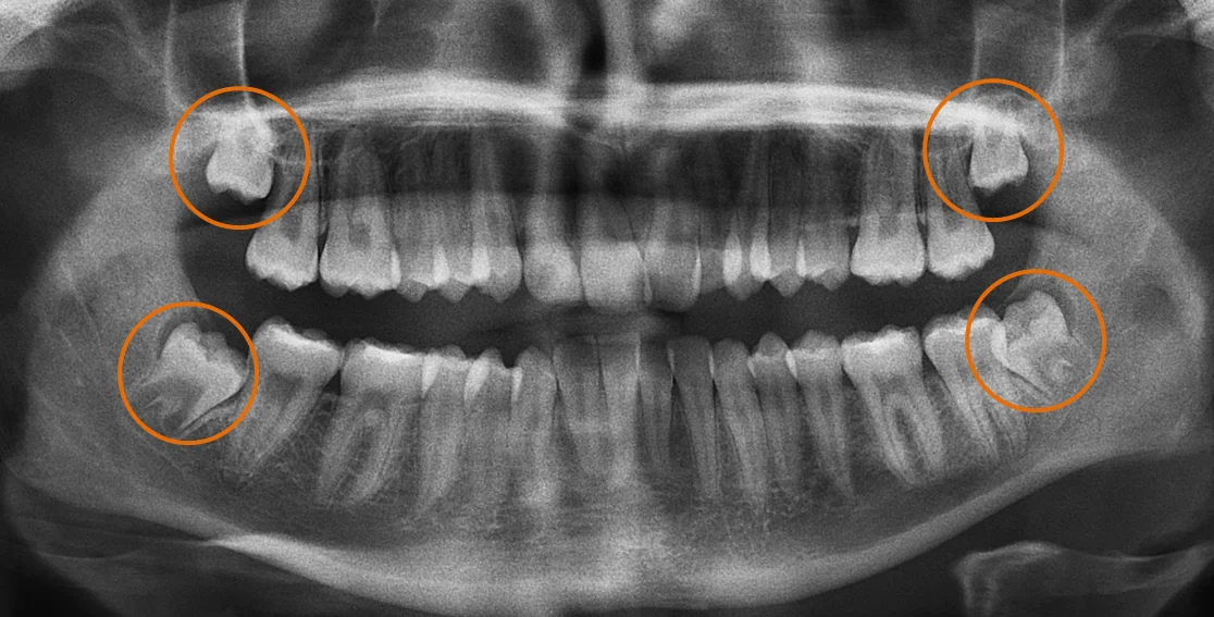
Panoramic X-rays: The Standard Tool
Panoramic x-rays create a single, flat image showing your entire jaw—all teeth, sinuses, and bone structure in one view.
For wisdom teeth, panoramic x-rays show:
- Whether wisdom teeth exist and their general position
- Approximate nerve canal location
- General proximity of roots to nerve
- Whether infection or cysts are present
What Panoramic X-rays Cannot Show
The critical limitation is dimensionality. Panoramic x-rays are two-dimensional projections of three-dimensional anatomy.
You cannot determine:
Whether the nerve runs in front of, behind, or between wisdom tooth roots. The 2D image collapses three-dimensional relationships—you see that nerve and root overlap on the x-ray, but don’t know if they’re touching or separated by millimeters with one in front of the other.
Exact root configuration. Roots might curve toward the nerve, wrap around it, or diverge away—impossible to determine from a single flat view.
Bone thickness between nerve and root. There might be substantial separation, paper-thin bone, or direct contact—indistinguishable on 2D imaging.
🔑 Key Takeaway: Standard x-rays aren’t inadequate due to poor quality—they’re dimensionally insufficient. Trying to remove wisdom teeth near nerves using only 2D images is like performing surgery while wearing a blindfold.
How CBCT 3D Scans Transform Wisdom Tooth Evaluation
CBCT (Cone Beam Computed Tomography) scanning creates three-dimensional images by capturing hundreds of x-ray views from different angles, reconstructing them into a 3D dataset.
What CBCT Reveals That Standard X-rays Cannot
For wisdom tooth evaluation, CBCT shows:
The nerve canal’s exact three-dimensional path through the jaw—not a flat line but an actual tube viewed from any angle.
Whether nerve and root actually contact or are separated. If they overlap on panoramic x-ray, CBCT reveals genuine contact versus structures at different depths.
Whether roots curve toward, away from, or around the nerve. Three-dimensional root anatomy becomes completely visible.
Bone thickness between nerve and root. CBCT measures precisely how much bone (if any) separates nerve from tooth.
Root canal anatomy and variations. Internal tooth structure becomes visible, revealing unusual anatomy that might complicate extraction.
Virtual Surgical Planning
Modern oral surgeons use CBCT data to virtually plan extraction before touching your mouth.
They can:
- Rotate 3D images to view wisdom teeth from any angle
- Measure exact distances from tooth to nerve at multiple points
- Identify the safest surgical approach based on anatomy
- Plan bone removal patterns that avoid nerve exposure
- Determine whether tooth sectioning is needed and where
- Assess whether coronectomy (removing only the crown) might be safer
This transforms extraction from reactive (“let’s carefully remove this and hope”) to proactive (“based on anatomy, here’s exactly how we’ll avoid the nerve 2mm behind the roots”).
At our CBCT imaging facility in Gandhinagar, advanced 3D dental CT scans provide exactly this level of anatomical detail.
When CBCT Is Essential for Wisdom Teeth
Not every wisdom tooth requires CBCT scanning. Understanding when 3D imaging is genuinely necessary helps you evaluate recommendations.
High-Risk Teeth Demanding CBCT
- Wisdom teeth showing panoramic signs of nerve proximity
- Horizontally or severely impacted teeth with roots pointing toward nerve
- Patients over 30 where root development is complete
- Previous failed extraction attempts
- Teeth with unusual root anatomy
- Upper wisdom teeth very close to sinuses
Lower-Risk Situations
- Simple, fully erupted wisdom teeth positioned normally
- Young patients with incompletely developed roots well above nerve
- Upper wisdom teeth with ample bone between roots and sinus
The decision should be individualized based on clinical examination, panoramic findings, patient age, and complexity assessment.
💡 Quick Tip: If cost concerns make you hesitant about CBCT, remember: fixing nerve damage complications costs far more than imaging. The question isn’t whether you can afford CBCT—it’s whether you can afford the complications working without it creates.
How CBCT Changes Surgical Approach
CBCT doesn’t just provide information—it changes what surgeons do during extraction.
Surgical Modifications Based on CBCT
Tooth sectioning patterns: CBCT shows precisely where to divide the tooth so sections can be removed individually, minimizing nerve exposure risk.
Bone removal planning: Three-dimensional anatomy reveals which bone must be removed and which should be preserved to protect the nerve.
Extraction sequence: CBCT might reveal that removing the crown first, then sectioning roots separately produces safer outcomes.
Coronectomy decisions: When CBCT shows roots intimately associated with nerve, surgeons might choose coronectomy (removing crown while leaving roots) rather than risking nerve damage.
Coronectomy: A CBCT-Guided Alternative
Coronectomy involves removing the wisdom tooth crown while deliberately leaving roots in the jaw.
For teeth with roots directly contacting or wrapping around the nerve, attempting complete removal carries high nerve injury risk. Coronectomy eliminates the problematic crown while avoiding nerve injury risk.
Studies show coronectomy success rates exceed 90% with very low complication rates. Roots often migrate away from the nerve over months as bone remodels.
However, coronectomy only makes sense when CBCT clearly demonstrates complete extraction carries unacceptable nerve risk.
Comparing Approaches: Standard vs. CBCT Imaging
| Factor | Panoramic X-ray Only | CBCT 3D Scan |
|---|---|---|
| Nerve Location | Approximate 2D position | Exact 3D pathway with millimeter precision |
| Root Anatomy | General shape in one plane | Complete 3D configuration |
| Nerve-Root Relationship | Overlap visible but contact unclear | Definitive: contact or separation measured |
| Surgical Planning | Reactive during surgery | Proactive—complete plan before incision |
| Nerve Injury Risk | Higher—incomplete knowledge | Lower—exact nerve location known |
| Coronectomy Decision | Difficult to determine | Clear indication based on anatomy |
| Patient Understanding | Limited explanation ability | Patients see 3D images, understand risks |
| Professional Standard | Outdated for high-risk cases | Current evidence-based approach |
What to Expect: CBCT-Guided Extraction Process
Step 1: Initial Evaluation
Oral surgeon examines wisdom teeth and reviews panoramic x-rays. If complexity or nerve proximity appears, CBCT is recommended.
Step 2: CBCT 3D Scanning
Scan takes 10-20 seconds—you remain still while machine rotates, capturing images. Processing creates 3D reconstruction for analysis.
Step 3: Detailed Surgical Planning
Surgeon spends 15-30 minutes analyzing CBCT:
- Tracing nerve canal through jaw
- Examining root anatomy from multiple angles
- Measuring nerve-root distances
- Planning specific surgical approach
Step 4: Pre-Surgical Consultation
Surgeon explains findings using 3D images—showing tooth position, nerve location, risks, and how planned approach minimizes those risks.
Step 5: The Extraction
Surgeon follows precise plan developed from CBCT analysis, constantly referencing mental map of nerve location throughout procedure.
At our facility, nitrous oxide sedation provides comfortable relaxation during extraction.
Step 6: Recovery
Recovery often proceeds more smoothly with CBCT-guided extractions because:
- More precise technique creates less tissue trauma
- Shorter surgical time reduces inflammation
- Complications were prevented rather than addressed reactively
For comprehensive dental care in Gandhinagar, CBCT-guided wisdom tooth extraction represents the modern standard.
Why Nova Dental Hospital for CBCT-Guided Wisdom Tooth Removal
At Nova Dental Hospital, Gandhinagar, wisdom tooth extraction is preceded by appropriate diagnostic imaging—including CBCT when anatomy demands it.
Our approach includes:
Advanced in-house CBCT 3D scanning providing complete anatomical visualization without external center referrals.
Expert analysis by Dr. Happy Patel with extensive experience interpreting CBCT for surgical planning.
Comprehensive surgical capabilities from simple erupted teeth to complex impacted cases requiring nerve preservation.
Same-day consultation and imaging when CBCT is indicated—no delays.
Transparent risk communication where we show actual 3D images, explain findings honestly, and discuss all options.
Comfortable sedation options including nitrous oxide for anxious patients.
Conveniently located near PDPU and Gift City, serving patients throughout Gandhinagar who expect modern care supported by advanced diagnostic technology.
Visit our Google Business profile to see patient experiences.
Frequently Asked Questions
Do all wisdom teeth need CBCT scans before removal?
No—CBCT is specifically indicated for wisdom teeth showing complexity or high nerve injury risk on standard x-rays. Simple, fully erupted wisdom teeth positioned normally don’t require CBCT. However, impacted wisdom teeth (especially horizontal or angled), teeth showing indirect signs of nerve proximity, wisdom teeth in patients over 30, and any teeth where the surgeon has anatomical concerns should be evaluated with CBCT. The decision should be individualized. When CBCT is recommended, it’s because your surgeon recognizes 2D imaging provides inadequate information for safe extraction in your case. Don’t decline recommended CBCT to save cost—the few thousand rupees for scanning protect you from nerve damage worth far more in medical expenses and quality of life impact.
What happens if CBCT shows my wisdom tooth root is touching the nerve?
If CBCT reveals direct root-nerve contact, several options exist depending on symptoms. For symptomatic teeth (pain, infection), coronectomy—removing crown while leaving roots—often provides the safest approach. This eliminates the problematic portion causing symptoms while avoiding nerve injury risk from root removal. Studies show coronectomy success rates exceeding 90% with roots often migrating from nerve over time. For asymptomatic teeth with concerning anatomy, watchful waiting might be safer than prophylactic extraction carrying high nerve risk. Some surgeons with advanced training might feel comfortable with complete extraction using careful technique, though accepting meaningful nerve injury risk. The key is CBCT allows informed decision-making—weighing symptom severity against complication risk.
How much does a wisdom tooth 3D x-ray cost?
CBCT scans for wisdom tooth evaluation typically cost ₹3,000-6,000 in Gandhinagar. Whether dental insurance covers CBCT depends on your plan—some cover diagnostic imaging when medically necessary, others have limitations. Most insurance recognizes CBCT for complex wisdom teeth is medically appropriate. Even without coverage, consider CBCT cost in context: the few thousand rupees protect your larger investment in extraction and prevent complications (nerve damage, additional surgeries) costing ₹50,000-100,000 to address. When CBCT is recommended based on panoramic findings suggesting nerve proximity, skipping it to save imaging fees is false economy—risking catastrophic complications to save relatively small amounts.
Can dental implants be placed safely without CBCT scans?
This question seems misplaced in wisdom tooth context, but addressing it: modern implant standards require 3D imaging for optimal safety. While experienced surgeons historically placed implants before CBCT existed, current evidence-based standards require 3D imaging. Studies show nerve injury rates, sinus perforation rates, and implant failure rates are significantly higher without CBCT guidance. Professional organizations state cross-sectional imaging should be considered for all implant cases. The question isn’t whether implants can ever succeed without CBCT—some do—but whether you want surgery planned with incomplete information when complete information is available.
How long do I have to wait between CBCT scan and wisdom tooth removal?
Wait time between CBCT and extraction depends on surgical scheduling, not medical requirements. CBCT scans remain valid 6-12 months unless significant changes occur. Some practices with in-house CBCT can perform imaging and extraction same day if scheduling allows. More commonly, scans are done at one appointment with extraction scheduled 1-2 weeks later after thorough planning. There’s no medical requirement to wait after CBCT—delay is purely logistical. If wisdom tooth is acutely infected, surgeon might prescribe antibiotics first, perform CBCT once acute symptoms subside, then schedule extraction. The key advantage of in-house CBCT is eliminating traditional multi-week delays from external imaging center referrals.
Related posts

Write a Comment
Recent Posts

Making Wisdom Tooth Removal Safer with CBCT Scans

X-rays Before Dental Implants: Using 3D Tech for Success


