
X-rays Before Dental Implants: Using 3D Tech for Success
X-rays Before Dental Implants: Using 3D Tech for Success
“My dentist says I need x-rays before getting dental implants. I just had regular x-rays at my last cleaning—aren’t those enough? Why do I need more imaging?”
This question reveals a common misunderstanding about dental imaging for implants. Not all x-rays are created equal, and what works perfectly for finding cavities is completely inadequate for planning implant surgery.
Here’s the reality that many patients don’t discover until they’re deep into implant treatment: The imaging done before your implant determines whether that implant succeeds or fails. It determines whether nerves are damaged. Whether the implant engages enough bone. Whether complications arise that could have been prevented.
Skipping proper pre-implant imaging isn’t just cutting corners—it’s gambling with your health and investment.
The difference between adequate imaging and inadequate imaging isn’t subtle. It’s the difference between placing an implant in the exact correct position with complete confidence versus hoping you didn’t drill too close to a nerve you couldn’t see. Between measuring precise bone dimensions versus estimating and hoping there’s enough. Between surgical precision and surgical guesswork.
This comprehensive guide explains exactly what imaging dental implants require and why, what standard x-rays can and cannot show, how 3D CBCT scanning transforms implant planning from approximate to exact, what information your surgeon gains from proper pre-implant imaging, and how this diagnostic step protects both your investment and your health.
Whether you’re just beginning to consider dental implants or you’re already scheduled for surgery, understanding the imaging foundation that successful implants require helps you evaluate whether you’re receiving appropriate care.
Understanding the Imaging Requirement: Why Implants Are Different
Before diving into specific imaging types, let’s establish why dental implants require more sophisticated diagnostic imaging than other dental procedures.
What Makes Implant Surgery Unique
Dental implants are surgical procedures involving permanent fixtures placed into your jawbone. Unlike fillings, crowns, or even tooth extractions, implant placement has zero tolerance for error in certain dimensions.
Consider what’s at stake:
Nerve damage from drilling too close to nerves causes permanent numbness—a devastating complication that proper imaging prevents.
Sinus perforation from placing upper implants too close to sinuses creates infections and potential implant failure—entirely avoidable with adequate imaging.
Inadequate bone engagement from misjudging bone dimensions leads to implant failure requiring removal and bone grafting—expensive and time-consuming to correct.
Incorrect implant positioning from insufficient pre-surgical visualization creates restorations that don’t align properly with opposing teeth—compromising both function and aesthetics.
Each of these complications is largely preventable through proper pre-surgical imaging. Each becomes significantly more likely when imaging is inadequate.
The Information Implant Surgery Demands
“What exactly does my implant surgeon need to know before placing the implant?”
Complete three-dimensional bone anatomy:
- Bone height at the implant site
- Bone width at every level where the implant will sit
- Bone quality and density
- Cortical (hard outer) versus trabecular (softer inner) bone distribution
Precise anatomical structure locations:
- Inferior alveolar nerve pathway in the lower jaw
- Mental foramen (nerve exit point) position
- Maxillary sinus floor contours in the upper jaw
- Incisive canal and nasopalatine nerve locations
- Any unusual anatomical variations or voids
Critical measurements:
- Exact distances from planned implant position to nerves
- Precise measurements from bone surface to sinus floor
- Available bone dimensions in all three axes
- Angulation possibilities given anatomical constraints
This information determines:
- Whether you’re even a candidate for implants without grafting
- What implant length and diameter are safe
- Optimal implant position and angulation
- Whether bone grafting or sinus lift procedures are needed first
- Surgical approach and technique selection
Standard 2D x-rays provide some of this information. CBCT 3D scans provide all of it. That’s why implant standards require 3D imaging.
💡 Quick Tip: If your dentist plans to place dental implants based on standard x-rays without CBCT 3D scanning, this is a serious red flag. Modern implant dentistry standards require 3D imaging—not because it’s impressive technology, but because it’s medically necessary for safe surgery.
🔑 Key Takeaway: Dental implants aren’t like other dental procedures where approximate information suffices. They require exact three-dimensional anatomical knowledge that only proper pre-surgical imaging provides. Cutting corners on imaging is cutting corners on your safety.
What Standard X-rays Show (and What They Miss)
Let’s examine what traditional dental x-rays actually reveal—and their critical limitations for implant planning.
Types of Standard Dental X-rays
Periapical x-rays show individual teeth and surrounding bone. These are the small x-rays where you bite down on a sensor.
What they show well: Tooth decay, root canal success, bone loss around specific teeth, abscess or infection at tooth roots.
What they cannot show: Bone width, three-dimensional nerve pathways, exact anatomical measurements needed for implants.
Bitewing x-rays show upper and lower teeth together, focusing on crowns and bone levels.
What they show well: Cavities between teeth, bone levels around multiple teeth, existing dental work quality.
What they cannot show: Root anatomy, bone dimensions beyond what’s visible in a single two-dimensional plane.
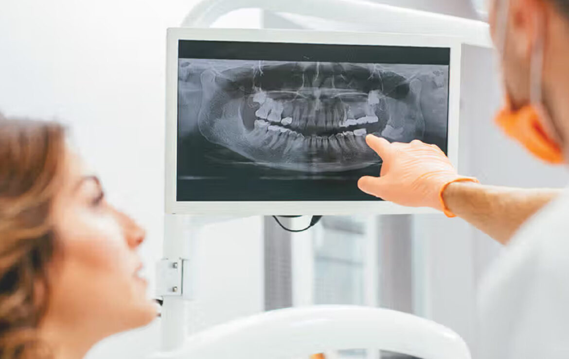
Panoramic x-rays (OPG) show the entire jaw, all teeth, sinuses, and TMJ joints in one large image.
What they show well: Overall dental and jaw structure, presence of all teeth including wisdom teeth, obvious pathology or problems, general bone levels.
What they cannot show: Precise measurements, bone width, exact nerve location, detailed anatomical relationships.
The Fundamental 2D Limitation
All standard x-rays share one critical limitation: they’re two-dimensional representations of three-dimensional anatomy.
Imagine trying to understand a building’s complete structure from a single photograph. You see the front facade—windows, doors, height. But you have no idea how deep the building is. Whether rooms are large or small. How the interior is laid out. Where staircases or support beams are located.
That’s exactly what 2D x-rays do for your jaw. They show one perspective—typically the side view. You see bone height. But bone width? Internal structure? Three-dimensional nerve pathways? These simply don’t exist in the two-dimensional image.
What This Means for Implant Planning
“Can’t my dentist just estimate bone width from the 2D x-ray?”
They can estimate. But estimation isn’t adequate for surgical placement of permanent implants.
Consider what happens with estimation:
If bone width is estimated at 10mm but is actually 7mm, the implant diameter selected might be too wide—resulting in bone perforation or inadequate engagement.
If the nerve appears to be 8mm below where you’re drilling but is actually 5mm below, you risk nerve damage.
If the sinus floor seems to be “plenty high” but closer examination reveals it’s much lower in that specific spot, you risk sinus perforation.
These aren’t theoretical risks—they’re complications that occur regularly when implants are planned from 2D imaging. Each represents a failure of diagnostics, not a failure of surgical skill.
For comprehensive CBCT 3D imaging in Gandhinagar, advanced diagnostic technology ensures your implant surgeon has complete anatomical information rather than estimates and approximations.
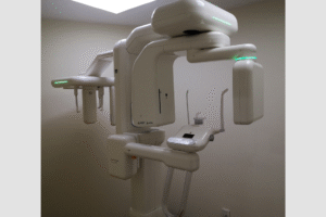
How CBCT 3D Scans Transform Implant Planning
CBCT (Cone Beam Computed Tomography) scanning creates three-dimensional images by taking hundreds of x-ray views from different angles, then reconstructing them into a complete 3D representation of your jaw.
What CBCT Actually Shows
Imagine being able to slice through your jaw like cutting a loaf of bread, examining each slice individually to see internal structure, then reassembling the slices to view from any angle.
That’s essentially what CBCT does digitally.
Your implant dentist can:
View cross-sections showing bone width at the exact location where the implant will be placed—not estimated, measured precisely.
Trace nerve pathways through the jaw in three dimensions, seeing exactly where nerves run and how close they come to the planned implant position.
Measure distances from the bone surface to the maxillary sinus with millimeter accuracy.
Assess bone quality through density measurements, identifying areas of dense cortical bone versus softer trabecular bone.
Identify anatomical variations like nerve pathway deviations, unusual blood vessel channels, or bone voids that would complicate surgery.
Plan implant positioning virtually by digitally placing the implant in the 3D scan before ever touching your mouth, optimizing position, angle, and depth.
The Virtual Implant Placement Advantage
Modern implant planning software allows your dentist to place virtual implants directly in your CBCT scan.
They can:
- Try different implant positions and angles
- Measure bone engagement at each possible position
- Verify adequate distance to nerves and sinuses
- Select optimal implant length and diameter
- Plan the final crown position and ensure implant placement supports it properly
- Create surgical guides based on the virtual plan
This virtual planning happens before surgery. By the time you’re in the surgical chair, every critical decision has been made based on complete anatomical knowledge.
Compare this to traditional approaches where many of these decisions were made during surgery based on what the surgeon encountered—a reactive rather than proactive approach.
Real-World Example: Upper Molar Implant
Let’s make this concrete with a specific example.
You need an implant to replace an upper back molar.
With standard 2D x-rays: Your dentist sees bone height appears adequate. The x-ray suggests the sinus is “probably high enough” for a standard implant. Bone width is unknown—assumed to be adequate based on clinical examination. Your dentist selects a standard 10mm length, 4.5mm diameter implant and proceeds with surgery hoping these dimensions work.
With CBCT 3D scanning: Cross-sectional views show bone width is 7mm (adequate for 4.5mm implant with safe margins). Precise measurement shows 11mm from bone crest to sinus floor (safe for 10mm implant with 1mm clearance). Bone density is measured and found to be good quality. Nerve pathway is mapped showing safe distance from planned position. Virtual implant placement confirms the 10mm × 4.5mm implant at 15-degree angle provides optimal bone engagement and positions the crown properly. Surgery proceeds with complete confidence because everything was measured, not estimated.
Which scenario would you prefer for your implant?
🔑 Key Takeaway: CBCT 3D scanning doesn’t just provide better images—it provides different information entirely. It shows dimensions and structures that are literally invisible in 2D x-rays, transforming implant planning from educated guessing to precise engineering.
What Your Implant Surgeon Learns from Pre-Implant Imaging
Let’s break down specifically how pre-implant CBCT imaging translates into surgical decisions and better outcomes.
Bone Dimension Assessment
The single most critical factor for implant success is adequate bone to support the implant.
From CBCT, your surgeon measures:
Bone height at the implant site with millimeter precision—determining maximum safe implant length.
Bone width at every level where the implant will sit—crucial because implants need bone surrounding them on all sides. A site might have 15mm of bone height but only 5mm of width—adequate height, inadequate width.
Available space between adjacent teeth or structures—ensuring the implant can be positioned without damaging neighboring teeth or compromising restoration space.
Bone quality variations—identifying areas of thinner cortical bone or softer trabecular bone that might affect initial stability or healing.
This dimensional assessment determines:
- Whether implant placement is possible without grafting
- Optimal implant diameter (wider is generally better but requires adequate bone)
- Maximum safe implant length
- Whether sinus lift or bone grafting is needed first
- Realistic timeline from diagnosis to completed restoration
Nerve Proximity Evaluation
Nerve damage is a catastrophic complication causing permanent numbness in the lip, chin, or gums.
CBCT allows precise nerve mapping:
The inferior alveolar nerve path is traced through the mandible in three dimensions. Your surgeon sees exactly where the nerve enters the jaw, how it courses through bone, where it’s closest to the surface, and where it exits (mental foramen).
Measurements are taken from the planned implant position to the nerve at multiple points, ensuring safe clearance (typically 2mm minimum, though surgeons often prefer 3-4mm margin for added safety).
Alternative positioning can be evaluated if the initial plan shows concerning nerve proximity—shifting the implant slightly forward or backward, changing the angle, or using a shorter implant to maintain safe distance.
This nerve evaluation prevents:
- Drilling directly into the nerve
- Placing the implant too close causing pressure on the nerve
- Post-surgical numbness from nerve trauma
- The devastating complication of permanent sensory loss
Sinus Relationship Planning
In the upper jaw, maxillary sinuses sit above the back teeth—sometimes quite close to where implants need to be placed.
CBCT reveals the complete sinus anatomy:
The three-dimensional sinus floor contours show whether the floor is flat, slopes, or has depressions dipping lower between teeth. Exact measurements from the bone crest to sinus floor determine available height for implant placement.
Sinus membrane thickness can be assessed (though detailed soft tissue is better visualized with medical CT, CBCT provides useful information). Anatomical variations like septa (dividing walls) within the sinus or unusual sinus pneumatization patterns are identified.
This sinus analysis determines:
- Whether standard implants can be placed with adequate clearance
- If sinus lift procedures are needed to create additional height
- What approach to sinus lifting is appropriate (lateral window vs crestal approach)
- Whether implants can be placed simultaneously with sinus lift or require staged surgery
- Risk factors for sinus perforation that demand extra surgical caution
For patients requiring dental implant treatment in Gandhinagar, comprehensive CBCT planning ensures surgical approaches account for all anatomical factors before beginning treatment.
Bone Grafting Need Identification
Often, the need for bone grafting is only identifiable through detailed 3D imaging.
CBCT reveals:
- Ridge width deficiencies not apparent clinically or on 2D x-rays
- Vertical bone loss requiring grafting for adequate height
- Fenestrations (windows) in the bone where the outer wall is missing
- Socket preservation needs if extraction and immediate implant are being considered
- Optimal grafting material volumes needed based on measured deficits
Identifying grafting needs during initial planning prevents:
- Aborted implant surgeries when inadequate bone is discovered mid-procedure
- Frustrated patients who thought treatment would be straightforward
- Extended treatment timelines from unplanned additional procedures
- Increased costs from procedures that should have been anticipated
💡 Quick Tip: If your dentist discovers mid-surgery that bone grafting is needed after planning was supposedly complete, this suggests inadequate pre-surgical imaging. Proper CBCT scanning should identify grafting needs during the planning phase, not during surgery.
When Standard X-rays Might Be Sufficient (Hint: Rarely)
“Are there any situations where standard x-rays without CBCT are adequate for implants?”
Professional guidelines state CBCT should be considered for all dental implant cases. However, let’s examine the rare exceptions where 2D imaging might theoretically suffice.
The Theoretical Exceptions
Ideal posterior implant sites with abundant bone visible on 2D x-rays, no history of bone loss or previous surgery, adequate clinical width on examination, and clear distance from nerves and sinuses might be placed with 2D imaging alone by very experienced surgeons.
However, even in these “ideal” cases, CBCT often reveals information that changes treatment planning—unexpected bone quality variations, anatomical peculiarities, or dimensions different from clinical estimates.
Why the Exceptions Don’t Matter
Here’s the practical reality: CBCT equipment is widely available, relatively inexpensive compared to implant surgery costs, and provides information that simply cannot be obtained any other way.
Choosing not to use CBCT when it’s available is choosing to work with incomplete information—there’s no medical justification for this when patient safety is paramount.
The question isn’t “when can we get away without CBCT?” but rather “why would we ever plan implant surgery without complete diagnostic information when that information is readily available?”
Modern implant dentistry standards are clear: CBCT imaging is appropriate for essentially all implant cases, not just complicated ones.
The Complete Pre-Implant Imaging Workflow
Understanding what happens from consultation through implant placement helps you appreciate how imaging fits into the complete treatment process.
Step 1: Initial Consultation and Clinical Examination
Your dentist examines your mouth, assesses general suitability for implants, takes medical history, and determines preliminary treatment approach.
Preliminary imaging (usually panoramic x-ray) provides overview of jaw anatomy and identifies obvious issues.
Decision point: Is the patient a reasonable implant candidate? If yes, proceed with detailed planning.
Step 2: CBCT 3D Scan
The diagnostic CBCT scan is performed, capturing complete three-dimensional jaw anatomy.
The scan itself takes 10-20 seconds. You remain still while the scanner rotates around your head capturing hundreds of images from different angles.
Processing creates the 3D reconstruction that your dentist will analyze.
Step 3: Detailed Treatment Planning
Your dentist spends 20-40 minutes (or more for complex cases) analyzing the CBCT scan:
- Measuring bone dimensions at planned implant sites
- Tracing nerve pathways and measuring distances
- Assessing sinus relationships and clearances
- Evaluating bone quality through density analysis
- Identifying any anatomical variations or concerns
Virtual implant placement in planning software optimizes position, angle, length, and diameter.
Treatment plan finalized: The surgical approach is determined, implant components are selected, any bone grafting needs are identified, and realistic timelines are established.
Step 4: Patient Consultation and Treatment Discussion
Your dentist shows you the 3D images (often rotating them so you see from different angles), explains findings and what they mean for your treatment, discusses any complications or additional procedures needed, answers questions about the planned approach, and reviews costs and timeline.
This consultation is far more informative when based on 3D imaging than when working from 2D estimates.
Step 5: Implant Surgery Execution
On surgery day, your surgeon references the CBCT images and planning throughout the procedure, verifies positioning as drilling progresses, and makes any necessary adjustments based on actual encountered conditions while staying within the parameters the pre-surgical planning established.
Many surgeons keep the CBCT images displayed on a screen in the surgical room, constantly referencing them during implant placement.
Step 6: Post-Surgical Verification
Sometimes a post-surgical x-ray or CBCT scan verifies implant position, ensures no complications occurred, and provides baseline imaging for future comparison.
This complete workflow shows how pre-implant CBCT imaging isn’t just “one more test”—it’s the foundation that all subsequent treatment decisions build upon.
🔑 Key Takeaway: Pre-implant imaging isn’t separate from treatment—it’s integral to it. The imaging determines what treatment is appropriate, what approaches are safe, and what outcomes are achievable. Skipping or inadequately performing this step undermines everything that follows.
Comparing Imaging Approaches: 2D vs. 3D for Implant Planning
| Factor | Standard 2D X-rays Only | CBCT 3D Scans |
|---|---|---|
| Bone Width Visibility | Not visible—must estimate | Precise measurements at all levels |
| Nerve Location | Approximate 2D position | Exact 3D pathway traced |
| Sinus Proximity | General idea of distance | Millimeter-precise measurements |
| Bone Quality Assessment | Subjective appearance | Quantitative density measurements |
| Virtual Planning | Not possible | Full virtual implant placement before surgery |
| Surgical Precision | Good surgeons work with limited info | Excellent—complete anatomical knowledge |
| Complication Risk | Higher—working with incomplete data | Lower—complications anticipated and avoided |
| Need for Bone Grafting | Often discovered during surgery | Identified during planning phase |
| Implant Sizing | Conservative estimates | Optimized for specific anatomy |
| Patient Confidence | “I hope this works” | “I know exactly what to expect” |
| Professional Standard | Outdated approach | Current evidence-based practice |
| Cost | Lower imaging fee, higher complication risk | Higher imaging fee, lower complication risk |
The Hidden Risks of Inadequate Pre-Implant Imaging
Let’s be direct about what can go wrong when implant planning relies on insufficient imaging.
Nerve Damage: The Permanent Complication
Inferior alveolar nerve injury during implant placement causes numbness in the lower lip, chin, and gums on the affected side.
This isn’t temporary post-surgical numbness—it’s permanent sensory loss. You might not feel hot or cold, might drool without realizing it, might bite your lip without noticing.
How often does this happen? Studies show nerve injury rates of 1-5% when implants are placed without CBCT guidance—far higher than the <0.5% rate with proper 3D imaging.
The tragedy is that every nerve injury from inadequate pre-surgical imaging was preventable. CBCT clearly shows nerve location. Drilling without this information is accepting unnecessary risk.
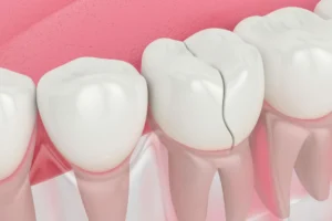
Implant Failure from Bone Inadequacy
Placing implants in insufficient bone is a recipe for failure.
If bone width is misjudged and the implant is wider than available bone, part of the implant ends up outside the bone (fenestration) or pushes through thin bone (perforation). These implants typically fail within months.
If bone quality is poor but not recognized pre-surgically, initial stability might be inadequate. The implant never integrates properly and must be removed.
These failures require:
- Implant removal
- Bone grafting to correct the deficiency
- Waiting 3-6 months for graft healing
- New implant placement attempt
- Additional costs often exceeding the original implant cost
All prevented by proper CBCT assessment before the first implant was placed.
Sinus Complications
Perforating the maxillary sinus during upper implant placement creates serious problems.
The sinus membrane tears, creating a pathway between your mouth (bacteria-filled) and sinus cavity (should be sterile). This can lead to:
- Chronic sinusitis requiring extended antibiotic treatment
- Implant failure necessitating removal
- Sinus membrane repair surgery
- Months of treatment delay while healing occurs
CBCT imaging shows exact sinus floor location, making these perforations almost entirely avoidable.
Financial Impact of Complications
Beyond the medical consequences, inadequate imaging creates financial burdens.
Consider the costs:
- Failed implant removal: ₹5,000-15,000
- Bone grafting to correct deficiency: ₹15,000-40,000
- Waiting period: Lost income, ongoing temporary solutions
- Replacement implant: ₹25,000-50,000
- Additional procedures for complications: Variable
Total cost of a failed implant often exceeds ₹50,000-100,000 beyond the original implant investment.
The few thousand rupees for CBCT imaging that could have prevented these complications seems quite reasonable in comparison.
For comprehensive dental care in Gandhinagar using appropriate diagnostic imaging, patients avoid the devastating complications that inadequate planning creates.
💡 Quick Tip: If cost concerns make you consider skipping CBCT imaging for implants, remember that fixing complications from inadequate imaging costs far more than proper diagnostic imaging upfront. The question isn’t whether you can afford CBCT—it’s whether you can afford the complications that working without it creates.
What to Expect During Your Pre-Implant CBCT Scan
If you’ve never had a CBCT scan, knowing what to expect helps you feel prepared.
Before the Scan
Preparation is minimal. You don’t need to fast, take medication, or do anything special before the scan.
Remove metal objects from your head and neck area—jewelry, glasses, hearing aids—as these create artifacts (distortions) in the images.
Inform your dentist if you’re pregnant or might be pregnant. While CBCT uses much less radiation than medical CT, dental procedures are typically postponed during pregnancy when possible.
During the Scan
You’ll sit or stand (depending on the machine type) with your chin resting on a support and your head positioned in the scanner.
Remain completely still for 10-20 seconds while the scanner rotates around your head. Any movement blurs the images.
The machine makes a gentle humming sound as it rotates. The process is completely painless—nothing touches your mouth or teeth.
The scan feels much shorter than it is because 20 seconds passes quickly when you’re focused on staying still.
After the Scan
You’re finished immediately. There’s no recovery, no waiting, no after-effects. You can drive, eat, work—normal activities resume immediately.
Processing takes a few minutes while the computer reconstructs your 3D images from the captured data.
Review happens either immediately (if your dentist analyzes images during the same appointment) or at a follow-up consultation (some practices prefer time to thoroughly analyze complex scans before discussing them with patients).
At our CBCT imaging facility in Gandhinagar, the entire scanning process is comfortable, quick, and efficient—providing the diagnostic information your implant surgery requires.
Choosing an Implant Provider: The Imaging Factor
“How much weight should I give to whether my dentist has CBCT capabilities when choosing where to get implants?”
Significant weight. Here’s why imaging capabilities reveal broader practice competence.
What In-House CBCT Signals
Practices that invest in CBCT equipment are telling you several things:
They’re serious about implant dentistry. CBCT costs hundreds of thousands of rupees. Practices make this investment because they do enough advanced procedures to justify it.
They understand modern standards. Current evidence-based implant dentistry requires 3D imaging. Practices aligned with modern standards have the equipment to meet those standards.
They value convenience. On-site imaging means treatment proceeds faster—consultation, imaging, and planning happen efficiently rather than spread across weeks and multiple locations.
They’ve invested in your success. The practice has committed significant resources to providing diagnostic capabilities that improve outcomes.
Questions to Ask
“Do you have CBCT imaging on-site, or will I need to go elsewhere for scans?”
In-house capabilities are strongly preferred for reasons discussed earlier—convenience, coordination, and treatment speed.
“Do you use CBCT imaging for all implant cases, or only complicated ones?”
The answer should be “all cases” or “essentially all cases.” Every implant deserves proper planning.
“Can you show me the 3D images and virtual implant placement during treatment planning?”
Being able to see and understand the imaging findings builds confidence and helps you participate in treatment decisions.
“How many implants have you placed, and what is your success rate?”
Experience matters. Success rates should be >95% for straightforward cases.
Red Flags
Be cautious if:
- The practice refers routine implant cases out for CBCT (suggests limited implant focus)
- The dentist says 2D x-rays are “usually sufficient” for implants
- They can’t clearly explain why CBCT is important
- Cost is cited as a reason to skip proper imaging
Your implant success depends on proper planning. Choosing a provider equipped for and committed to appropriate diagnostic imaging is choosing someone committed to your success.
Why Nova Dental Hospital for Your Implant Imaging and Treatment
At Nova Dental Hospital, Gandhinagar, proper diagnostic imaging is non-negotiable for implant success.
Our approach to pre-implant imaging includes:
Advanced in-house CBCT 3D scanning providing complete three-dimensional visualization of your jaw anatomy without referring you to external imaging centers.
Comprehensive scan analysis by Dr. Happy Patel with extensive implant experience, ensuring imaging findings translate into optimal surgical planning.
Virtual implant planning software allowing digital implant placement in your 3D scan before surgery, optimizing position, angle, and sizing based on your unique anatomy.
Same-day consultation and imaging for efficient treatment timelines—no weeks of delay waiting for external imaging appointments and results.
Transparent treatment planning where you see the actual 3D images, understand the findings, and participate in decisions about your treatment approach.
Comprehensive implant capabilities from simple single implants to complex full mouth rehabilitation—all supported by the diagnostic foundation proper CBCT imaging provides.
Conveniently located near PDPU and Gift City, we serve patients throughout Gandhinagar who expect modern implant dentistry supported by appropriate diagnostic technology.
Visit our Google Business profile to see what patients say about experiencing properly planned implant treatment.
Frequently Asked Questions
Do I really need x-rays before dental implants if I just had regular x-rays at my cleaning?
Yes—dental implants require CBCT 3D scans specifically, not the standard x-rays taken during routine cleanings. Regular dental x-rays (periapicals, bitewings, or even panoramic x-rays) are excellent for finding cavities, checking existing dental work, and monitoring gum disease. However, they cannot provide the three-dimensional anatomical information that implant surgery requires. Think of it this way: Your routine x-rays are like photographs showing what’s visible from one angle. CBCT 3D scans are like a complete architectural blueprint showing internal structure from every angle. Both have their purposes, but implant surgery needs the blueprint, not just the photograph. The fact that you recently had dental x-rays for other purposes doesn’t eliminate the need for specialized pre-implant CBCT imaging. These are different diagnostic tools for different purposes, and proper implant planning requires the information only CBCT provides.
How much does a CBCT scan cost before dental implants, and is it included in the implant price?
CBCT scan costs typically range from ₹3,000-6,000 as a separate imaging fee in most practices. Whether this is included in overall implant treatment costs varies by practice—some include imaging in package pricing while others charge separately. When evaluating costs, consider the true value: the few thousand rupees for CBCT imaging protects your much larger investment in the implants themselves (often ₹25,000-50,000 per implant) by preventing complications that could necessitate implant removal, bone grafting, and replacement at costs exceeding ₹50,000-100,000. Additionally, factor in that some practices refer you to external imaging centers where you might pay similar scan fees plus travel costs, time off work, and treatment delays—while in-house CBCT provides better value through integrated care. When discussing treatment costs with your dentist, ask specifically what imaging is included versus charged separately, and understand that proper diagnostic imaging isn’t an “optional extra” but rather a medical necessity for safe implant placement.
Can dental implants be placed safely without CBCT scans?
While experienced surgeons historically placed implants before CBCT technology existed, modern evidence-based standards require 3D imaging for optimal safety and success. The question isn’t whether implants can ever succeed without CBCT—some certainly do—but rather whether you want your surgery planned with incomplete information when complete information is readily available. Studies consistently show that nerve injury rates, sinus perforation rates, and implant failure rates are all significantly higher when implants are placed without CBCT guidance compared to with proper 3D imaging. Professional organizations including the American Academy of Oral and Maxillofacial Radiology state that cross-sectional imaging should be considered for all dental implant cases. Just as you wouldn’t want orthopedic surgery performed without MRI when that diagnostic tool is available, dental implants shouldn’t be placed without CBCT when this technology exists and is appropriate. The relevant question isn’t “can it be done without CBCT?” but rather “why would I accept higher complication risks by skipping diagnostic imaging that clearly improves outcomes?”
What if my dentist says my case is “simple” and doesn’t need 3D imaging?
Ask your dentist to explain specifically why they consider CBCT unnecessary for your particular situation. Legitimate reasons might include: recent CBCT scans already exist from another provider that adequately show the anatomy, you’re only in preliminary consultation stage and detailed imaging will occur before actual surgery, or there are specific medical contraindications to additional radiation exposure. However, if reasoning is “I can tell from clinical examination that bone is adequate” or “I’ve placed hundreds of implants without CBCT,” these aren’t medically sound justifications. What seems “simple” on 2D imaging or clinical examination often reveals complexities on CBCT—unexpected bone narrowing, nerve pathway variations, or anatomical factors that change the treatment approach. Current professional standards recommend CBCT for essentially all implant cases, not just obviously complicated ones. Many complications arise specifically in “simple” cases where pre-surgical imaging would have revealed factors that weren’t apparent through clinical examination alone. You’re entitled to request appropriate diagnostic imaging, and dentists committed to evidence-based practice should welcome rather than resist this request.
How long is a CBCT scan valid—do I need a new one if implant surgery is delayed?
CBCT scans typically remain valid for 6-12 months if no significant changes have occurred in your mouth. Factors that might necessitate new imaging include: substantial time passing since the original scan (over 12 months), intervening dental procedures that changed anatomy (extractions, bone grafting, other surgeries), new dental problems developing (infections, tooth loss) since the original imaging, or original scans of insufficient quality or missing critical views. If you had CBCT imaging at one practice but are now consulting with a different implant provider, bring copies of your scans—most dentists can review existing imaging rather than repeating scans unnecessarily. However, some situations warrant new imaging even if previous scans exist: if treatment planning requires different views than what was captured originally, if image quality was suboptimal, or if significant anatomy changes have occurred. During consultation, your new dentist can review existing imaging and determine whether it’s adequate or if updated scans are necessary. Having recent CBCT available typically saves both time and money, though proper treatment planning never compromises on having the imaging information surgery requires.
Related posts

Write a Comment
Recent Posts

X-rays Before Dental Implants: Using 3D Tech for Success


