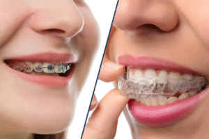
Why 2D X-rays Aren’t Enough for Dental Implants: The Critical Role of 3D Imaging
Why 2D X-rays Aren’t Enough for Dental Implants: The Critical Role of 3D Imaging
“My dentist says I need a 3D scan before getting dental implants, but they already took regular x-rays. Isn’t that enough? Why do I need more imaging?”
This is one of the most common questions patients ask when planning dental implant treatment. And it’s an excellent question because it touches on something fundamental about implant success that many people don’t realize.
Here’s the uncomfortable truth: placing dental implants using only standard 2D x-rays is like performing surgery while wearing a blindfold. You might get lucky. You might place the implant perfectly. But you’re making critical decisions—decisions that affect nerve function, sinus integrity, and implant longevity—based on incomplete information.
The difference between 2D and 3D imaging isn’t just technical sophistication. It’s the difference between guessing and knowing. Between hoping the bone is thick enough and measuring it precisely. Between estimating where nerves run and seeing their exact path. Between crossing your fingers and having complete confidence.
This comprehensive guide explains why 2D x-rays fundamentally cannot provide the information dental implant surgery requires, what 3D CBCT scans reveal that 2D images miss entirely, how this technology prevents complications that once seemed unavoidable, and why choosing a practice with advanced 3D imaging capabilities matters more than you might think.
Whether you’re planning your first implant or trying to understand why your consultation recommended 3D scanning, this information helps you appreciate why the best implant dentists won’t proceed with surgery until they’ve seen your jaw anatomy in three dimensions.
Understanding the Fundamental Limitation of 2D X-rays
Before exploring what 3D imaging provides, it’s crucial to understand what standard x-rays simply cannot show—not because they’re poor quality, but because of physics.
How 2D X-rays Actually Work
Standard dental x-rays create flat, two-dimensional images by passing x-ray beams through your jaw and capturing them on a sensor or film behind your teeth.
Think of it like taking a photograph of a building. The photo shows the front of the building clearly—you can see windows, doors, and architectural details. But you have no idea how deep the building is. You can’t see what’s behind the front wall. You don’t know if there’s a single room or fifty rooms inside.
That’s exactly what 2D x-rays do for your jaw. They show one view—typically the side view—of your teeth and bone. You see height clearly. But width? Depth? Internal bone structure? Those dimensions simply don’t exist in the image because the x-ray collapsed three-dimensional anatomy into a two-dimensional picture.
What This Means for Dental Implants
“Why does that matter for dental implants?”
Because dental implant surgery happens in three dimensions. The implant is a cylinder being placed into bone—it has length, width, and depth. The implant must:
- Fit within available bone width (not just height)
- Avoid nerves that run through the jaw
- Stay far enough from sinus cavities above back teeth
- Be surrounded by adequate bone on all sides for stability
None of these critical factors can be accurately assessed from a flat, 2D image. A 2D x-ray might show that bone height is adequate, but tell you nothing about whether there’s enough width. It might suggest a nerve is “somewhere in that area” without showing its precise three-dimensional location.
💡 Quick Tip: If your dentist says they can place implants based on 2D x-rays alone without 3D imaging, that’s a significant red flag. Modern implant dentistry standards require 3D imaging for safe, predictable outcomes—not because it’s fancy technology, but because it’s medically necessary.
🔑 Key Takeaway: 2D x-rays aren’t inadequate because of poor quality—they’re inadequate because they’re dimensionally insufficient. Trying to place dental implants from 2D images alone is like trying to park a car using only your side mirrors. You need to see all dimensions to do it safely.
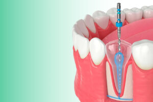
What 3D CBCT Scans Reveal That 2D X-rays Cannot
CBCT (Cone Beam Computed Tomography) 3D scans create complete three-dimensional reconstructions of your jaw anatomy. Instead of a single flat image, you get hundreds of “slices” that, when combined, show your jaw from every conceivable angle.
Bone Width and Thickness
The most critical measurement for implant success is bone width at the precise location where the implant will be placed.
On a 2D x-ray, bone width is invisible. You see height—you can tell if bone extends vertically where you need it. But you have no idea if that bone is 3mm wide (too narrow for most implants) or 12mm wide (plenty of room).
3D CBCT scans measure bone width at every point. You can see cross-sections showing exactly how wide the bone is at the top, middle, and bottom of where the implant will go. If the bone narrows in the middle (which happens frequently), the 3D scan reveals this—while a 2D x-ray would miss it entirely.
Why this matters: Placing an implant in bone that’s too narrow leads to implant failure. The implant either fails to integrate (osseointegrate) properly, or it does integrate initially but fails months or years later when it can’t handle chewing forces. 3D imaging prevents this by showing exactly how much bone exists before surgery even begins.
Nerve Location with Precision
The inferior alveolar nerve runs through your lower jaw, providing sensation to your lower lip, chin, and gums. Damage to this nerve during implant surgery causes numbness—sometimes permanent.
On a 2D x-ray, you can see that the nerve is “somewhere in the lower jaw.” You might estimate it’s “probably about 10mm from the top of the bone.” But that’s an estimate based on a two-dimensional projection of a three-dimensional structure.
3D CBCT scans trace the exact three-dimensional path of the nerve. You see precisely where it enters the jaw, how it curves through the bone, and where it’s closest to potential implant sites. Measurements aren’t estimates—they’re exact distances from the planned implant position to the nerve at every point along its course.
Why this matters: Drilling too close to the nerve can cause permanent numbness—a devastating complication that’s almost entirely preventable with proper 3D imaging. Studies show nerve injury occurs far more frequently when implants are placed without CBCT guidance.
Sinus Proximity and Anatomy
The maxillary sinuses sit above your upper back teeth. When placing implants in this area, you must stay far enough below the sinus floor to avoid perforating into the sinus cavity.
On a 2D x-ray, the sinus appears as a dark area above the teeth. You can see it’s “up there somewhere.” But exact distances? The three-dimensional shape of the sinus floor? Whether the floor dips down between teeth or stays level? Invisible.
3D CBCT scans map the complete sinus anatomy in three dimensions. You see the sinus floor’s exact contours, measure precise distances from bone crest to sinus, and identify areas where the sinus dips lower (making implant placement trickier).
Why this matters: Accidentally perforating the sinus during implant surgery creates serious complications—sinus infections, implant failure, need for sinus membrane repair. Proper 3D imaging shows exactly how much clearance exists, eliminating guesswork entirely.
For comprehensive CBCT 3D imaging in Gandhinagar, advanced cone beam technology provides the complete anatomical visualization that safe implant surgery requires.
Bone Quality Assessment
Not all bone is created equal. Dense, compact bone provides better implant stability than soft, porous bone.
On a 2D x-ray, bone density is suggested by how white (radiopaque) the area appears. But this is qualitative and imprecise—you’re essentially eyeballing whether bone “looks dense.”
3D CBCT scans provide quantitative bone density measurements through Hounsfield units (similar to what medical CT scans use). You can see exactly which areas have dense cortical bone versus softer trabecular bone, allowing precise implant site selection and appropriate implant design choices.
Why this matters: Placing implants in poor-quality bone without recognizing the density issue leads to initial instability, longer healing times, or outright failure. Knowing bone quality in advance allows your surgeon to modify technique, choose appropriate implant designs, or recommend bone grafting when needed.
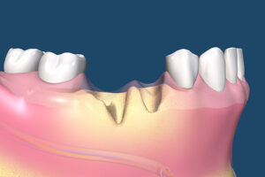
Complex Anatomical Variations
Every jaw is unique. Some people have thin bone ridges. Others have unusual nerve pathways. Some have large nutrient canals running through their bone. Some have concavities or undercuts invisible on 2D images.
3D CBCT scans reveal all of these anatomical variations that might complicate implant surgery. What looks straightforward on a 2D x-ray might reveal on 3D imaging to be far more complex—or occasionally, easier than expected.
Why this matters: Surprises during surgery are bad. Finding unexpected bone deficits or anatomical variations mid-procedure forces on-the-fly decision-making and increases complication risks. Complete pre-surgical knowledge from 3D imaging allows thorough planning before the first incision.
Real Complications That 2D Imaging Cannot Prevent
Understanding theoretical limitations is one thing. Seeing real-world consequences makes the importance concrete.
Nerve Damage from Misjudged Proximity
“My implant was placed fine, but afterward, my lip went numb. My dentist said it was ‘just bad luck.'”
It’s not bad luck—it’s inadequate imaging. When implants are placed too close to the inferior alveolar nerve based on 2D estimations rather than 3D measurements, permanent numbness can result.
This isn’t rare. Studies show that nerve injuries during implant surgery occur significantly more frequently when procedures are performed without CBCT guidance. The numbness might be partial or complete. It might resolve over months or be permanent. Either way, it was preventable.
Sinus Perforation
When the implant drill or the implant itself perforates the sinus membrane, you’ve introduced a connection between your mouth (full of bacteria) and your sinus cavity (supposed to be sterile). This can lead to:
- Chronic sinusitis requiring extended antibiotic treatment
- Implant failure and need for removal
- Sinus membrane repair surgery
- Months of delayed treatment while healing occurs
3D imaging shows the exact distance to the sinus floor, making these perforations almost entirely avoidable. When they occur despite CBCT imaging, it’s typically in very thin bone where the risk was known and accepted—not an unexpected complication from inadequate planning.
Implant Failure from Insufficient Bone
“My implant failed after six months. The dentist said ‘sometimes they just don’t take.'”
Sometimes implants fail despite perfect placement. But many “failures” result from placing implants in bone that was too narrow, too soft, or too thin—factors a 3D scan would have revealed before surgery.
When an implant is placed in bone that’s narrower than the implant diameter, part of the implant ends up outside the bone (called “fenestration”) or pushes through the thin bone wall (“perforation”). These implants rarely succeed long-term.
3D imaging prevents these placement errors by showing exact bone dimensions before surgery. If bone is insufficient, bone grafting can be done first—adding months to treatment but preventing implant failure.
Complications from Unknown Anatomical Variations
Unusual nerve pathways, large nutrient canals, or unexpected bone voids occasionally exist in locations where implants are planned. On a 2D x-ray, these might be invisible or appear as vague shadows easily dismissed.
When the surgeon encounters these unexpectedly during surgery, the procedure becomes more complicated. Sometimes the implant position must be changed mid-surgery. Sometimes the implant can’t be placed at all without aborting the procedure and replanning.
3D CBCT scans identify these variations before surgery, allowing thorough pre-operative planning that accounts for anatomical challenges from the start.
For expert dental implant treatment in Gandhinagar using advanced 3D CBCT planning, patients benefit from implant success rates that comprehensive imaging supports.
🔑 Key Takeaway: These complications aren’t just theoretical risks—they happen regularly when implants are placed without adequate 3D imaging. Every complication described above is dramatically less likely when proper CBCT scanning guides the surgery.
How 3D Imaging Changes the Implant Planning Process
CBCT 3D scans don’t just provide better images—they transform the entire planning process from educated guessing to precise engineering.
Virtual Implant Placement Before Surgery
Using specialized software, your dentist can virtually place the implant in the 3D scan before touching your mouth. They can:
- Rotate the implant to find the optimal angle
- Measure exactly how long the implant can be without hitting nerves or sinuses
- Determine the largest diameter implant the bone can accommodate
- Identify the ideal position for optimal crown emergence and aesthetics
- Verify adequate bone surrounds the implant from all sides
This virtual planning happens weeks before surgery. By the time you’re in the surgical chair, every critical decision has been made based on complete anatomical knowledge.
Compare this to traditional approaches where the surgeon makes many of these decisions during surgery based on what they encounter. Virtual planning eliminates real-time guesswork.
Guided Surgery Using 3D Planning
Some dentists take CBCT planning a step further with guided surgery. The virtual implant placement from the 3D scan is converted into a physical surgical guide—a custom-made template that fits over your teeth and guides the drill to the exact planned position.
With guided surgery:
- The implant goes exactly where the 3D plan shows it should
- Angle and depth are controlled by the guide, not estimated
- Surgical time often decreases significantly
- Accuracy improves dramatically
Not all implant cases require guided surgery. Simple single implants often don’t need this level of precision. But complex cases—multiple implants, full-arch rehabilitation, implants in tight anatomical spaces—benefit enormously from guided approaches that CBCT imaging makes possible.
Identifying the Need for Bone Grafting Before Surgery
“I thought I was coming in for an implant, but they said I need bone grafting first. Why wasn’t this clear earlier?”
When planning is done from 2D x-rays, bone deficiency often isn’t apparent until surgery begins. The surgeon makes the first cut and discovers “there’s not enough bone here—we need to graft.”
This means:
- Today’s surgery is aborted or modified
- You need a second surgery months later
- Treatment timeline extends unexpectedly
- Costs increase
3D CBCT scans reveal bone deficiencies during the planning phase, before you’re numbed, before the first incision, before time and money are invested in an approach that won’t work. If grafting is needed, it’s planned from the beginning—no surprises, no aborted surgeries, no frustrated patients wondering why this wasn’t caught earlier.
At our dental clinic in Gandhinagar, comprehensive 3D imaging ensures treatment planning accounts for all anatomical factors before you commit to surgery.
💡 Quick Tip: If your dentist discovers mid-surgery that bone grafting is needed after planning was supposedly complete, this suggests the planning was inadequate—likely based on 2D imaging that couldn’t reveal the deficiency. This shouldn’t happen with proper 3D CBCT planning.
Comparing 2D vs. 3D Imaging for Implant Planning
| Critical Factor | 2D X-rays | 3D CBCT Scans |
|---|---|---|
| Bone Width Measurement | Cannot see bone width—dimension is collapsed | Precise measurements at every level—see exact width in cross-sections |
| Nerve Location | Approximate position based on 2D projection | Exact three-dimensional nerve pathway traced with precise distances |
| Sinus Distance | General proximity visible, exact distance unknown | Millimeter-precise measurements; see complete sinus anatomy |
| Bone Quality | Subjective assessment by appearance | Quantitative density measurements (Hounsfield units) |
| Anatomical Variations | Many variations invisible or dismissed as shadows | All variations visible in three dimensions |
| Implant Angle Planning | Estimated during surgery | Precisely planned virtually before surgery |
| Implant Length Selection | Conservative estimates to avoid complications | Exact maximum safe length calculated |
| Bone Grafting Need | Often discovered during surgery | Identified during planning phase |
| Guided Surgery Capability | Not possible without 3D data | Enables surgical guide fabrication |
| Complication Risk | Higher—nerve injury, sinus perforation, implant failure | Dramatically lower with complete anatomical knowledge |
| Surgical Surprises | Common—unexpected findings mid-procedure | Rare—anatomy fully known before first incision |
| Overall Approach | Educated guessing based on incomplete information | Precise engineering based on complete data |
The Safety and Radiation Question
“Isn’t a CT scan a lot more radiation than regular x-rays? Is it safe?”
This is a legitimate concern that deserves a straightforward answer.
CBCT Radiation Compared to Other Imaging
CBCT scans do use more radiation than a single 2D dental x-ray. However, the radiation dose is significantly lower than medical CT scans—often 10 to 20 times less radiation than a full medical CT.
To put this in perspective:
- Single 2D dental x-ray: Minimal radiation exposure
- Full mouth series of dental x-rays: Moderate exposure (comparable to CBCT)
- Single CBCT scan: Moderate exposure (similar to full mouth x-rays)
- Medical CT scan: Significantly higher exposure
The key question isn’t “is there radiation?” but “is the benefit worth the minimal risk?”
Risk-Benefit Analysis for Implant Planning
For dental implant surgery, the answer is clearly yes. The complications prevented by proper 3D imaging—nerve damage, sinus perforation, implant failure—carry far more risk than the minimal radiation exposure from a single CBCT scan.
Think of it this way: Would you rather accept minimal radiation exposure from one CBCT scan, or risk permanent numbness from nerve damage that 2D imaging couldn’t prevent? Would you rather have one CBCT scan, or multiple surgeries because bone grafting need wasn’t identified until mid-surgery?
Modern CBCT technology also minimizes radiation through focused beam areas (scanning only the specific region needed, not your entire head), optimized exposure protocols, and high-efficiency sensors requiring less radiation for quality images.
When CBCT Is Medically Necessary
For dental implant planning, CBCT isn’t optional extra imaging—it’s medically necessary. The American Academy of Oral and Maxillofacial Radiology states that cross-sectional imaging (CBCT) should be considered for all dental implant cases.
This isn’t technology for technology’s sake. It’s the appropriate diagnostic tool for the procedure being performed. Just as you’d expect an orthopedic surgeon to order an MRI before knee surgery rather than relying on x-rays alone, dental implants require 3D imaging to be placed safely and predictably.
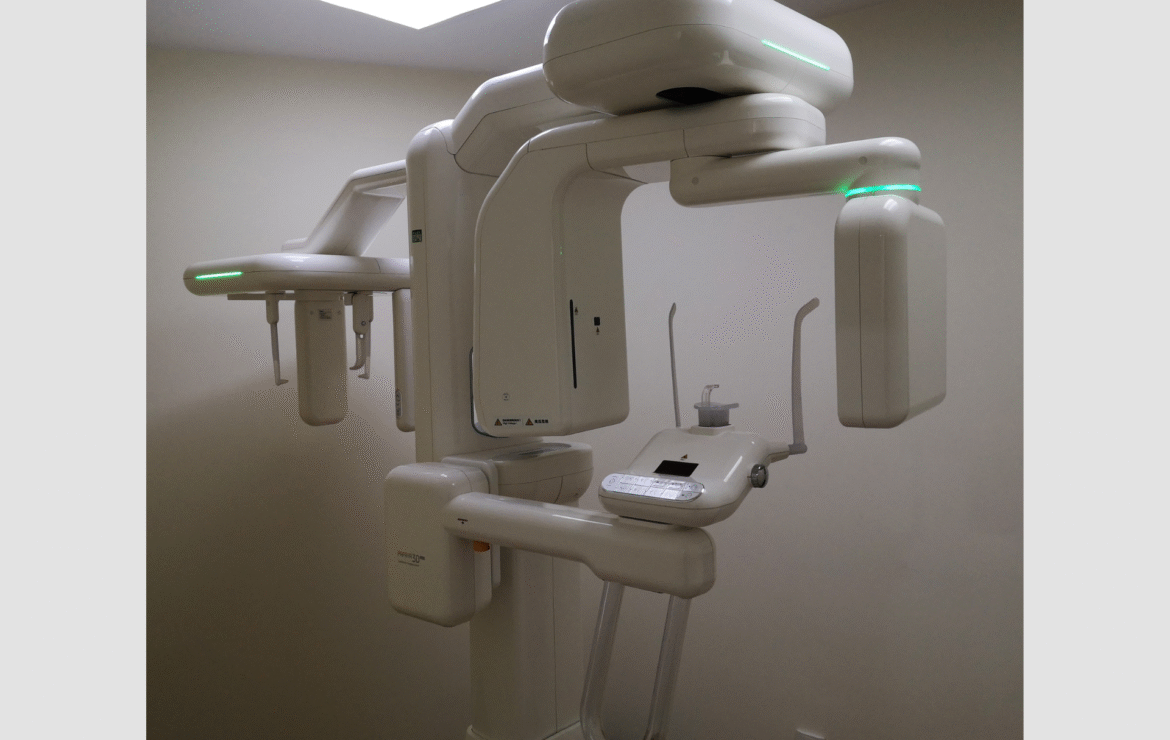
What to Expect During a CBCT Scan
If you’ve never had a CBCT scan, knowing what to expect helps you feel prepared.
The Scanning Process
The scan itself takes about 10-20 seconds. You sit or stand (depending on the machine) while the scanner rotates around your head capturing images. You need to remain still during this brief time, similar to holding still for a photograph.
Most CBCT machines have a chin rest or forehead rest to help you stay positioned correctly. The machine makes a gentle humming sound as it rotates. There’s no enclosed tube like medical CT scans—most CBCT machines are open and don’t trigger claustrophobia.
The process is completely painless. No injection, no contrast dye, nothing touching your teeth or mouth. Just sitting or standing still for a few seconds while images are captured.
After the Scan
Your dentist receives hundreds of image slices showing your jaw from every angle. Using specialized software, they examine these images to assess bone dimensions, locate nerves, measure sinus distances, and plan implant placement.
This analysis might take 20-30 minutes of careful review. Some dentists perform this analysis during your consultation appointment; others review the scans before your next visit so they can discuss findings and treatment planning with you.
You’ll typically see the images yourself during treatment planning—your dentist can show you the bone dimensions, point out the nerve pathways, and explain why implants can or cannot be placed in specific locations.
For state-of-the-art CBCT 3D dental imaging in Gandhinagar near PDPU and Gift City, modern equipment and experienced interpretation ensure you receive the diagnostic information your implant surgery requires.
💡 Quick Tip: Some dental practices don’t have CBCT scanners in-house and send patients to imaging centers. While this works, having CBCT available at your implant dentist’s office is more convenient—scanning can be done the same day as consultation, and your dentist can review images immediately with you present.
When 2D Imaging Might Still Be Used
Does this mean 2D x-rays are useless? Absolutely not.
Appropriate Uses for 2D Imaging
2D x-rays remain excellent for:
- Routine dental examinations and cavity detection
- Monitoring existing dental work
- Evaluating gum disease bone loss
- Checking root canal treatment quality
- General screening for dental problems
For these purposes, standard x-rays provide the information needed without the additional radiation of 3D imaging.
The issue isn’t that 2D x-rays are bad—it’s that they’re insufficient for implant planning specifically. Using the right imaging for the right purpose is what matters.
Simple Implant Cases Might Use Both
Even with CBCT available, many dentists take 2D x-rays first. These initial films provide a general overview and help determine if you’re even a reasonable implant candidate before investing in more advanced imaging.
For instance, a 2D x-ray might show that bone height is completely inadequate—so inadequate that implants clearly aren’t viable without extensive grafting. In this case, the 2D x-ray provided useful screening information before CBCT was necessary.
Think of 2D x-rays as screening and 3D CBCT as diagnostic planning for implants. Both have roles, but only 3D provides the information implant surgery requires.
How to Evaluate Your Implant Dentist’s Imaging Capabilities
“I’m choosing between dentists for my implant. Should their imaging capabilities influence my decision?”
Absolutely yes. The technology available significantly affects the quality and safety of your treatment.
Questions to Ask
Does your practice have a CBCT scanner on-site, or do I need to go elsewhere for 3D imaging?
Having CBCT in-house is more convenient and allows immediate integration of imaging into treatment planning. Sending patients elsewhere isn’t a deal-breaker, but in-house capabilities suggest a practice committed to advanced implant dentistry.
Do you routinely use CBCT for dental implant planning?
The answer should be yes. If a dentist says “we can get by with regular x-rays for most cases,” that’s concerning. Modern implant standards call for 3D imaging in essentially all cases.
Can you show me the 3D images and measurements during treatment planning?
Being able to see the actual scans—the bone measurements, nerve locations, and planned implant position—helps you understand the treatment plan and feel confident about the approach.
Do you use implant planning software with the CBCT data?
Software that allows virtual implant placement in the 3D scan represents a higher level of planning precision than simply viewing CBCT images without digital planning tools.
Red Flags
Be cautious if:
- The dentist downplays the need for 3D imaging (“we’ll just use regular x-rays”)
- CBCT is available but not recommended for your case without clear explanation
- The practice has never invested in 3D imaging capabilities despite offering implants
- Cost is cited as the reason to skip CBCT (proper imaging is a cost of doing implants safely)
Your implant’s success depends on accurate planning. Choosing a dentist committed to using appropriate diagnostic tools is choosing someone committed to your best outcomes.
🔑 Key Takeaway: Implant dentistry is advancing rapidly. Practices still relying primarily on 2D imaging for implant planning are using outdated approaches that increase complication risks. Modern implant dentistry requires—and patients deserve—3D imaging guidance.
The Cost Consideration: Is 3D Imaging Worth It?
“CBCT scans cost more than regular x-rays. Is it really worth the extra expense?”
Let’s examine this honestly.
The Direct Costs
Yes, CBCT imaging costs more than 2D x-rays. The equipment itself costs hundreds of thousands of dollars. The specialized software requires ongoing licensing fees. The training to interpret 3D images properly takes time and expertise.
These costs are passed to patients through imaging fees that exceed standard x-ray fees.
The True Cost-Benefit Analysis
However, consider the costs of complications CBCT prevents:
Nerve damage requiring specialist evaluation, possible nerve microsurgery, and dealing with permanent numbness—immeasurable cost to quality of life, plus thousands in medical expenses.
Sinus perforation requiring sinus repair surgery, antibiotics, implant removal and replacement—several thousand dollars and months of extended treatment.
Implant failure from placement in inadequate bone requiring implant removal, bone grafting, waiting for healing, new implant—often $5,000-10,000 in additional treatment.
Bone grafting discovered mid-surgery requiring aborted procedure, separate grafting surgery, and delayed implant placement—lost surgical time, additional anesthesia, extended treatment timeline.
Against these potential costs, the few hundred dollars for CBCT imaging becomes a modest investment in preventing expenses that dwarf the imaging fee.
What Proper Imaging Actually Buys You
Beyond preventing complications, CBCT imaging buys:
Confidence knowing your surgery is planned based on complete information, not guesswork.
Predictability with precise planning eliminating most surgical surprises.
Success as implants placed with 3D guidance have higher long-term success rates.
Time efficiency through surgical planning that prevents mid-procedure changes and aborted surgeries.
Peace of mind knowing your dentist saw exactly what they were working with before the first incision.
Is that worth a few hundred dollars? For most patients facing implant surgery, absolutely yes.
For comprehensive dental care in Gandhinagar using advanced diagnostic technology, the investment in proper imaging supports outcomes that cost-cutting on diagnostics undermines.
Why Nova Dental Hospital in Gandhinagar for Your Implant Imaging
Not all CBCT scans are equal. The technology matters, but so does the expertise interpreting the images.
At Nova Dental Hospital, Gandhinagar, our approach to implant imaging combines:
Advanced CBCT technology providing high-resolution 3D scans with minimal radiation exposure and focused scanning of only the necessary areas.
Expert image interpretation from Dr. Happy Patel with extensive implant dentistry experience understanding exactly what to look for in 3D scans and how to translate imaging findings into surgical planning.
Integrated treatment planning using specialized software that allows virtual implant placement directly in your 3D scan before surgery begins.
Convenient on-site imaging so your consultation, CBCT scan, and treatment planning happen in one location without traveling to separate imaging facilities.
Comprehensive implant capabilities from simple single implants to complex full mouth rehabilitation—all supported by the diagnostic precision CBCT imaging provides.
Transparent communication where we show you the actual 3D images and explain exactly what we see, what we’re planning, and why, so you’re fully informed about your treatment.
Located conveniently near PDPU and Gift City, we serve patients throughout Gandhinagar who seek implant dentistry supported by advanced diagnostic imaging and surgical expertise.
Visit our Google Business profile to see what patients say about their implant experiences with our practice.
Frequently Asked Questions
Why can’t my dentist just place the implant and see what they find during surgery?
Because “seeing what you find” during surgery often means finding problems you can’t fix on the spot. If the surgeon discovers bone is too narrow mid-procedure, they can’t suddenly create wider bone—the surgery must be aborted and bone grafting scheduled for months later. If they discover the nerve is closer than expected, they either risk nerve damage by proceeding or change the implant position without proper planning for how that affects the final crown. These mid-surgery discoveries create complications, delays, and suboptimal outcomes that proper 3D imaging prevents entirely. Modern surgical standards in all medical fields involve knowing the anatomy completely before making the first incision—dental implant surgery is no different. The “see what we find” approach was how implants were placed decades ago when better imaging didn’t exist. Today, it’s simply poor practice that increases risks unnecessarily.
Can CBCT scans show everything, or are there still unknowns?
CBCT scans show anatomical structures with remarkable precision, but some factors remain variable until surgery. Bone density measurements are very accurate, but the exact quality of bone integration during healing involves biological processes imaging can’t fully predict. Soft tissue thickness above bone is visible but can change slightly between imaging and surgery. The patient’s immune response, healing capacity, and individual biological factors affect outcomes in ways no imaging can determine. However, these limitations are minor compared to the massive unknowns of working without 3D imaging. With CBCT, we know bone dimensions precisely, see nerve locations exactly, measure sinus distances accurately, and identify anatomical variations clearly—all factors that are completely unknown or poorly estimated from 2D x-rays alone. Perfect knowledge isn’t achievable in medicine, but CBCT provides the information we need to plan and execute implant surgery with high predictability and safety.
I’ve heard some dentists say they can place implants successfully using just regular x-rays based on experience. Is that true?
Experienced implant surgeons can sometimes place implants successfully using 2D imaging, but “sometimes” isn’t the standard you want for your surgical care. Yes, dentists placed thousands of successful implants before CBCT technology existed—using educated guessing, conservative approaches, and accepting higher complication rates as inevitable. The question isn’t whether implants can ever succeed without 3D imaging—it’s whether you want your surgery based on incomplete information when complete information is available. Would you want a surgeon operating on you using guesswork and experience alone when MRI or CT scans could show exactly what they’re dealing with? Experience doesn’t create X-ray vision through bone—it just makes educated guessing slightly better. Modern patients deserve more than educated guessing, and modern technology provides it. Dentists who still rely primarily on 2D imaging for implants are using outdated approaches that increase risks unnecessarily.
Does the radiation from a CBCT scan cause cancer?
The radiation exposure from a single dental CBCT scan is minimal and the cancer risk is vanishingly small—far lower than many everyday radiation exposures. To put it in perspective, a CBCT scan typically exposes you to about the same radiation as 2-4 months of natural background radiation from simply living on Earth, or roughly equivalent to a cross-country airplane flight. Medical research studying radiation risks has not identified increased cancer rates from dental CBCT scans at these low exposure levels. The theoretical cancer risk from one CBCT scan is estimated at less than 1 in 100,000—compared to the very real risks of nerve damage (roughly 1-5 in 100 without 3D imaging) or implant failure (10-20 in 100 without proper planning). Risk exists in all medical procedures, but the relevant question is whether benefits outweigh risks. For dental implant planning, the benefits of CBCT (preventing serious complications, enabling precise planning, improving success rates) vastly outweigh the minimal theoretical cancer risk from brief radiation exposure.
My dentist said I don’t need a CBCT scan because my case is “simple.” Should I insist on one anyway?
Ask your dentist to explain specifically why 3D imaging isn’t necessary for your particular case. Legitimate reasons might include: you’re getting a very simple single implant in an area where bone is obviously abundant, recent CBCT scans already exist from another procedure, or you’re only in the consultation phase and CBCT will be done before actual surgery. However, if the reasoning is “I can tell from 2D x-rays that there’s enough bone” or “I’ve done this many times without CBCT,” those aren’t adequate justifications. Any implant placement involves risks best managed through complete anatomical knowledge, which only 3D imaging provides. Current professional standards recommend CBCT for essentially all implant cases. You’re entitled to request 3D imaging for any implant you’re receiving, and dentists committed to optimal patient care should welcome rather than resist that request. If your dentist is defensive about the suggestion, consider whether that’s someone you want placing permanent implants in your jaw.
Related posts