
Digital 3D Scan for Braces and Smile Design
Digital 3D Scan for Braces and Smile Design
Before the Braces Go On, the Scan Comes First
There is a moment early in every orthodontic or smile design consultation when the conversation shifts from talking about your goals to actually measuring where you are starting from. That is the diagnostic phase — and in modern dentistry, it begins with a scan.
The days of sitting with a tray full of cold, chalky impression material in your mouth are largely behind us. Today, the scan for braces and smile design is digital, precise, and far more informative than traditional plaster moulds ever were. Whether your dentist is planning orthodontic braces, clear aligners, or a full smile design with veneers, the imaging taken at this stage shapes every clinical decision that follows.
This blog explains what types of digital scans are used in orthodontic and cosmetic dentistry, what each one reveals, how they work together, and why the quality of your pre-treatment imaging directly influences the quality of your result.
🔑 Key Takeaways
- A scan for braces today typically means a digital intraoral scan, an OPG, and in complex cases, a CBCT — each capturing different diagnostic information.
- 3D teeth scans have replaced physical dental moulds for most orthodontic and cosmetic cases — with greater accuracy, faster turnaround, and better patient comfort.
- CBCT scans are used selectively in orthodontics — primarily for impacted teeth, skeletal discrepancies, and complex surgical cases.
- Smile design relies on digital surface scans combined with photographs and facial analysis to create a preview of the final result before treatment begins.
- All three imaging modalities are available in-house at Nova Dental Hospital — integrated into a single diagnostic workflow.
What Is a Scan for Braces — and Why Has It Replaced Moulds?
If you had braces a decade or two ago, you likely remember impressions: a U-shaped tray loaded with a thick, setting material pressed firmly into your mouth and held there for several minutes while you tried not to gag. The resulting plaster models were then used to study your bite, plan tooth movements, and fabricate appliances.
That process worked — but it had real limitations. Plaster models could warp or break. They had to be physically stored for years. Sharing them between specialists meant shipping them. And the accuracy, while adequate, was inherently limited by the physical properties of impression materials.
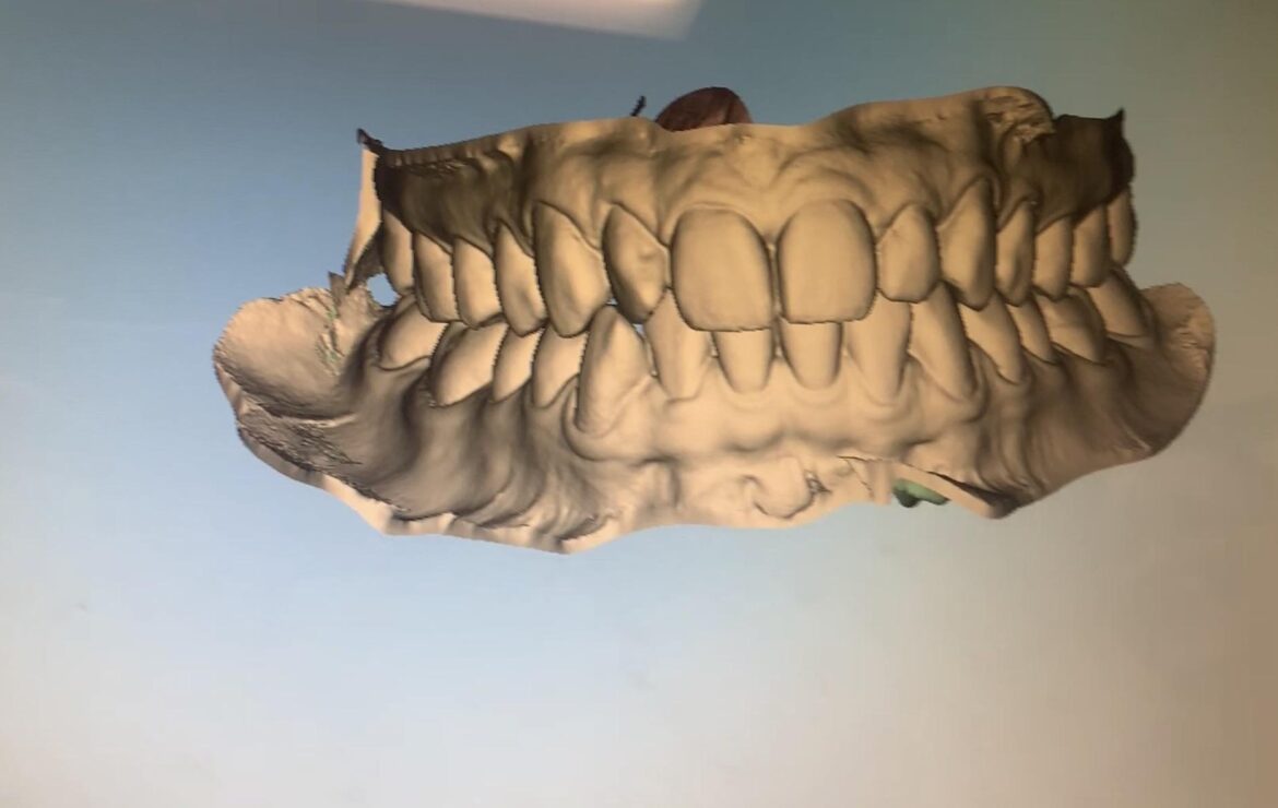
A digital 3D teeth scan eliminates all of these limitations. Using a small, handheld intraoral scanner — a wand-like device with a camera at its tip — your dentist captures thousands of images per second as it is moved gently around the mouth. Software stitches these images together in real time, creating a highly accurate three-dimensional digital model of your teeth, gums, and bite.
The whole process typically takes three to five minutes. There is nothing to swallow, nothing that sets in your mouth, and no discomfort. The digital model is available immediately on screen — and can be shared with orthodontic laboratories, aligner manufacturers, or specialist colleagues in seconds.
What an Intraoral Scan Captures
An intraoral 3D scan records:
- Crown morphology — the exact shape, size, and surface detail of every visible tooth
- Dental arch form — the curvature and width of both the upper and lower arches
- Occlusion — how the upper and lower teeth meet when the jaws are closed
- Spacing and crowding — precise millimetre measurements of gaps and overlaps
- Gum contour — the gingival line and soft tissue profile around each tooth
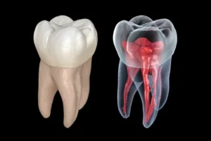
What an intraoral scan does not capture is everything below the gumline — root position, bone levels, nerve pathways, and jaw structure. For that, additional imaging is needed. This is why a scan for braces in a thorough clinical setting is never just one type of scan.
The OPG: The Panoramic Foundation of Every Orthodontic Plan
Before any orthodontic treatment begins, your dentist needs to see far more than the crowns of your teeth. An OPG (Orthopantomogram) — the full-jaw panoramic X-ray — provides the wider diagnostic picture that an intraoral scan cannot: root lengths and angulations, bone levels, the presence and position of unerupted or impacted teeth, the development status of wisdom teeth, and the symmetry of the jaw joints.
For orthodontic planning, the OPG answers several clinically critical questions:
- Are there any unerupted teeth, and if so, where are they positioned relative to the roots of adjacent teeth?
- Are the roots of teeth at risk of resorption if moved in a particular direction?
- Is there adequate bone support for planned tooth movements?
- Are wisdom teeth present, and will they need to be removed before or during orthodontic treatment?
- Is there any underlying pathology — cysts, infections, or bone irregularities — that would affect the treatment plan?
An OPG is considered a mandatory pre-treatment record in orthodontics. It is taken alongside the intraoral scan, clinical photographs, and in some cases a cephalometric (lateral skull) X-ray — all of which together form the complete diagnostic record from which your treatment plan is built.
✅ Quick Tip: The Orthodontic Diagnostic Record
- A complete orthodontic diagnostic record typically includes: intraoral 3D scan, OPG, lateral cephalometric X-ray, clinical photographs (front, side, and intraoral), and a clinical examination of jaw joints and facial symmetry.
- Each element answers a different question — no single scan replaces the others.
- At Nova Dental Hospital, all of these records are taken in-house and reviewed by your treating specialist before a treatment plan is presented.
When Is a CBCT Scan Used in Orthodontics and Smile Design?
A CBCT scan is not part of every orthodontic or cosmetic case — and it should not be. As covered in our earlier guide on CBCT vs OPG imaging, the 3D scan is reserved for cases where two-dimensional imaging leaves important clinical questions unanswered. In orthodontics and smile design, there are specific, well-defined scenarios where CBCT adds diagnostic value that genuinely changes the treatment plan.
Impacted Canines and Unerupted Teeth
Impacted upper canines are one of the most common indications for CBCT in orthodontics. An OPG can tell you that an impacted canine is present, but it cannot tell you precisely where that tooth sits in three dimensions — whether it is palatally or buccally positioned, how close its root apex is to adjacent teeth, and what angle and force will be needed to bring it into alignment through orthodontic traction.
A CBCT scan resolves all of this. It maps the impacted tooth’s exact position, identifies any root resorption it may already be causing to neighbouring teeth, and allows the orthodontist and oral surgeon to plan the surgical exposure and traction mechanics with precision before the first incision is made.
Skeletal Discrepancies and Jaw Asymmetry
When the upper and lower jaws are significantly misaligned — either in the front-to-back, vertical, or lateral dimension — orthodontic tooth movement alone may not achieve a functional or aesthetic result. These cases often involve a combination of orthodontics and jaw surgery (orthognathic surgery).
CBCT imaging in these cases provides the three-dimensional skeletal analysis needed to:
- Quantify the degree of jaw discrepancy in all three planes
- Assess the symmetry of the jaw joints (condyles) and the surrounding bone
- Plan the surgical movements required to reposition the jaws
- Create surgical splints and guides from the 3D data
Airway Assessment in Orthodontics
For patients with crowded arches requiring expansion, or those with potential sleep-disordered breathing, CBCT allows volumetric measurement of the airway. This is increasingly relevant in both adult and paediatric orthodontics, where the relationship between arch development and airway space has become an important clinical consideration.
Root Proximity and Alveolar Bone Boundaries
In cases where teeth are severely crowded and roots are in close proximity, CBCT helps the orthodontist map the available bone envelope through which teeth can safely move. This prevents root fenestrations (where a root tip exits the bone boundary) and reduces the risk of root resorption during treatment.
✅ Quick Tip: CBCT in Orthodontics — When It Is and Is Not Needed
- CBCT IS indicated: impacted canines or incisors, orthognathic surgery planning, severe skeletal asymmetry, complex root proximity, airway assessment.
- CBCT is NOT routinely needed: standard crowding and spacing cases, routine overbite or overjet correction, most clear aligner cases without impaction.
- If your orthodontist recommends a CBCT, ask which specific clinical question it is being used to answer — a good clinician will always explain.
- For a deeper understanding of radiation doses involved in dental CBCT, our radiation safety guide explains the numbers plainly.
3D Teeth Scans and Clear Aligner Treatment
Clear aligner systems — including Invisalign and its equivalents — are built almost entirely around 3D digital scan data. The treatment works by fabricating a series of precisely graduated aligner trays, each one moving teeth by fractions of a millimetre toward their planned final positions. That level of precision is only achievable when the starting point is captured with the accuracy of a digital intraoral scan.
Physical impressions are still accepted by some aligner manufacturers, but digital scans have become the clear standard — they are more accurate, faster to process, and eliminate the dimensional changes that can occur when physical impressions are poured into stone models and then digitised. Most leading aligner manufacturers now prefer or require digital scan submissions.
From Scan to Simulation: The ClinCheck and Treatment Preview
Once the digital scan is submitted, software generates a virtual treatment simulation — a step-by-step animation showing how each tooth is planned to move across the entire treatment sequence, from the first aligner to the last. At Nova Dental Hospital, this simulation is reviewed with the patient before treatment begins, allowing you to see a digital preview of your projected end result.
This is one of the most significant advantages of digital scanning in clear aligner treatment: the treatment plan becomes visible and reviewable before a single aligner is manufactured. Adjustments can be made at the digital planning stage — repositioning the final tooth positions, modifying the rate of movement, or adjusting the planned outcome — without any physical cost.
Refinement Scans During Treatment
Clear aligner treatment often requires one or more refinement scans midway through treatment. As teeth move, the actual positions may diverge slightly from the predicted digital plan — particularly in complex movements or in patients where compliance with wear time has been inconsistent. A fresh intraoral scan at the refinement stage captures the current tooth positions accurately, allowing a new set of aligners to be fabricated from the updated data.
For patients undergoing Invisalign or clear aligner treatment at Nova Dental Hospital, these refinement scans are taken in-house — keeping the entire treatment process integrated and efficient.
3D Scanning in Smile Design: From Digital Wax-Up to Final Veneers
Smile design is a process of planning aesthetic dental changes — whether through porcelain veneers, composite bonding, crown reshaping, or a combination — to achieve a harmonious, proportionate, and natural-looking result. The digital scan sits at the very centre of modern smile design workflows.
The Digital Smile Design Process
A smile design at Nova Dental Hospital begins with a multi-dimensional diagnostic record that combines:
- Intraoral 3D scan — capturing tooth shapes, proportions, and their relationship to the gumline
- Clinical photographs — full face, profile, lip at rest, lip retracted, and close-up tooth views
- Facial analysis — assessing midline, smile arc, tooth display at rest and when smiling, facial symmetry, and lip-to-tooth relationship
- Functional assessment — how the teeth meet, any wear patterns, and jaw joint position
These elements are combined in digital smile design software to create a treatment simulation — overlaying proposed tooth shapes and positions onto your actual facial photographs, so you can see and approve the planned changes before any tooth preparation begins.
The Digital Wax-Up and Mock-Up
From the digital scan and smile design plan, a digital wax-up is produced — a three-dimensional model showing the proposed final tooth shapes. This is used by the dental laboratory to fabricate the porcelain veneers or crowns, and it also forms the basis of a temporary mock-up that can be placed directly onto your teeth in the clinic.
The mock-up is a critical step. It allows you to see, feel, and speak with the proposed new tooth shapes before any irreversible preparation takes place. You can assess how the changes look in natural light, in photographs, and when you smile in conversation — not just in a dental chair. Adjustments to shape, length, or proportions can be made at this stage at no cost.
This workflow — scan, design, digital wax-up, mock-up, approval, fabrication — is the modern standard for smile design and veneer treatment at Nova Dental Hospital. It means no surprises, no guesswork, and a result that has been collaboratively agreed before the first drill is used.
When CBCT Adds Value in Smile Design
For straightforward veneer cases where the underlying teeth and bone are healthy, a CBCT is not required. The intraoral scan, OPG, and clinical photographs provide sufficient information. However, CBCT becomes relevant in smile design cases that involve:
- Implant placement as part of smile restoration — where bone mapping is essential
- Gum contouring surgery — where bone crest levels and tooth root lengths need 3D assessment
- Combined orthodontic and restorative treatment — particularly where impacted teeth are involved
- Full mouth rehabilitation — where the relationship between jaw position, bone, and planned restorations requires a complete 3D picture
For patients whose smile design involves any surgical or implant component, our in-house CBCT and OPG imaging facility ensures the full diagnostic picture is available in a single visit.
Digital Scanning in Orthodontics and Smile Design — A Complete Overview
The table below summarises which scanning technologies are used across different treatment types and what each contributes to the clinical plan:
| Treatment | Intraoral 3D Scan | OPG | CBCT | Primary Purpose |
| Dental Braces (standard) | ✅ Required | ✅ Required | ⬜ Usually not needed | Tooth positions, arch form, root overview |
| Braces with impacted teeth | ✅ Required | ✅ Required | ✅ Required | 3D localisation of impaction |
| Clear Aligners / Invisalign | ✅ Required | ✅ Required | ⬜ Unless complex | Digital treatment planning and tray fabrication |
| Orthognathic Surgery Planning | ✅ Required | ✅ Required | ✅ Required | Full skeletal 3D analysis |
| Veneers / Smile Design | ✅ Required | ✅ Required | ⬜ If implants or surgery involved | Digital wax-up, proportions, lab communication |
| Full Mouth Rehabilitation | ✅ Required | ✅ Required | ✅ Usually required | Complete restorative and surgical planning |
| Implant as part of smile plan | ✅ Required | ✅ Required | ✅ Required | Bone mapping, angulation, nerve safety |
Physical Impressions vs. Digital Scans: Why the Shift Matters
It is worth being specific about why the move from physical impressions to digital scans represents a genuine clinical improvement — not just a convenience upgrade.
| Factor | Physical Impression | Digital Intraoral Scan |
| Patient comfort | Gag reflex common, material taste unpleasant | Comfortable, nothing to swallow or set |
| Accuracy | Can warp, bubble, or distort on removal | Sub-0.1mm accuracy in modern systems |
| Processing time | Poured in stone, 24–48 hours | Immediate — model available in minutes |
| Storage | Physical models require storage space for years | Cloud-stored digitally, accessible instantly |
| Lab communication | Physical shipping, risk of damage in transit | Instant digital file transfer to laboratory |
| Aligner fabrication | Scan of physical model introduces second-stage error | Direct digital submission — no intermediate step |
| Refinements | Requires new physical impression | Quick rescan, typically 2–3 minutes |
| Reproducibility | Variable between clinicians | Consistent and measurable across scans |
The accuracy advantage is particularly significant for clear aligner treatment, where cumulative errors in tooth position across thirty or forty aligner stages can lead to significant divergence from the planned outcome. Starting from the most accurate digital record reduces this risk substantially.
What to Expect at Your Scan Appointment at Nova Dental Hospital
Many patients arrive for their first orthodontic or smile design consultation uncertain about what the diagnostic appointment involves. Here is a clear picture of what typically happens:
Intraoral Digital Scan
Your dentist or clinical team member will pass a small, handheld scanner wand gently around your mouth, moving it systematically across all tooth surfaces. You will be able to see the 3D model building in real time on the screen beside you — most patients find this genuinely interesting. The scan takes approximately three to five minutes for a full arch, or around two minutes for a single arch.
There is no preparation required, no numbing, and nothing that will cause any physical sensation beyond the slight presence of the wand in your mouth.
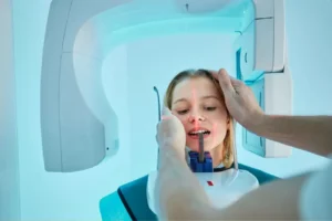
OPG X-ray
You will stand at the OPG unit and bite gently on a small, sterile chin rest. The machine’s arm rotates around your head in approximately 15 to 20 seconds, capturing the full panoramic image. The image appears on our digital system immediately.
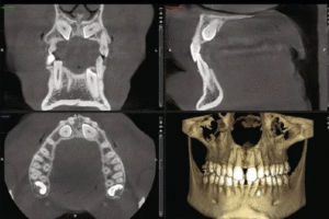
CBCT Scan (When Applicable)
If a CBCT scan has been indicated for your case, you will be positioned in the CBCT unit with your head gently stabilised. The rotating arm completes a 360° sweep in 10 to 40 seconds depending on the field of view selected. A lead apron is provided as standard. The 3D reconstruction is processed immediately and reviewed by your specialist in the same appointment.
Photographs and Facial Analysis
For smile design cases, a standardised set of clinical photographs is taken — typically including a full-face front view, profile, relaxed lip position, smiling, and close-up intraoral views. These are used alongside the digital scan to build the facial analysis and design the proposed changes in proper proportion to your unique facial features.
Consultation and Plan Review
Because all imaging at Nova Dental Hospital is in-house and digital, your treating specialist can review the full diagnostic record in the same appointment. Rather than waiting days for external scan results, your clinical photographs, digital scan model, OPG, and any CBCT data are all available on screen during your consultation — allowing a detailed, informed conversation about your treatment options before you leave the clinic.
✅ Quick Tip: Preparing for Your Diagnostic Appointment
- Brush and floss before your appointment — a clean mouth produces a cleaner intraoral scan.
- Remove any lip or tongue piercings beforehand — metal objects interfere with both the intraoral scanner and X-ray imaging.
- Bring a list of any previous orthodontic treatment — previous retainers, extractions, or braces — so your dentist has a complete history.
- If you have had recent dental X-rays at another clinic, request a copy to bring with you — this may reduce the need for additional imaging.
- Come with questions about your smile goals. The diagnostic appointment is as much about understanding your priorities as it is about taking records.
Frequently Asked Questions
FAQ 1: Do I need a separate scan for braces and another for clear aligners, or is it the same scan?
The same intraoral digital scan is used whether you are planning braces or clear aligners. The scan data can be used for both — for orthodontic analysis by your clinician, and for digital submission to an aligner manufacturer if clear aligners are the chosen treatment. The only difference is that clear aligner treatment relies entirely on the digital scan for fabrication, whereas braces require the scan primarily for planning, with the physical brackets bonded directly to teeth.
FAQ 2: Is the 3D teeth scan the same as a CBCT?
No — they are different technologies that capture different information. An intraoral 3D teeth scan uses optical camera technology to map the visible surfaces of your teeth and gums — it involves no radiation and captures only what is above the gumline. A CBCT scan uses X-ray technology to generate a three-dimensional image of teeth, roots, bone, and nerves — capturing everything below the gumline as well. In a thorough orthodontic or smile design workup, both types of scan may be used, each providing information the other cannot.
FAQ 3: Will I be able to see a preview of my smile before treatment begins?
Yes — for smile design cases at Nova Dental Hospital, a digital simulation is created from your scan and photographs that shows the proposed changes overlaid onto your actual face. For clear aligner cases, a step-by-step virtual treatment animation shows how your teeth are planned to move across the entire treatment sequence. These previews are reviewed with you before any treatment is confirmed, and adjustments can be made at the digital planning stage.
FAQ 4: How accurate are digital scans compared to traditional impressions for braces?
Modern intraoral scanners achieve sub-0.1mm accuracy across a full arch — consistently more accurate than physical impressions, which are subject to distortion from material properties, removal technique, and the subsequent pouring process. For clear aligner fabrication specifically, studies have shown that directly scanned digital models result in better aligner fit and more predictable tooth movement compared to digitised physical impressions. The accuracy of the starting record directly influences the precision of the final result.
FAQ 5: Does Nova Dental Hospital use digital scanning for all orthodontic and smile design cases?
Yes. Nova Dental Hospital uses digital intraoral scanning as standard for all orthodontic and smile design cases. Combined with in-house OPG and, where indicated, CBCT imaging, our diagnostic workflow is fully digital from the first scan to the final treatment plan. Patients who would like to share their experience are also welcome to leave a review on our Google Business Profile — it helps other patients in Gandhinagar understand what modern dental care looks like.
🔑 Key Takeaways
- A scan for braces in a modern dental clinic involves an intraoral 3D scan, an OPG, and in select cases a CBCT — each contributing different diagnostic information.
- Digital 3D teeth scans have replaced physical impressions as the clinical standard — offering greater accuracy, immediate availability, and seamless lab integration.
- CBCT is used selectively in orthodontics: primarily for impacted teeth, orthognathic surgery, and cases where root or bone boundaries need 3D mapping.
- Clear aligner treatment is built on digital scan data — accuracy of the starting scan directly influences the precision and predictability of tooth movement.
- Smile design workflows combine intraoral scans with facial photographs to produce digital wax-ups, treatment simulations, and physical mock-ups before any irreversible treatment begins.
- At Nova Dental Hospital, all scanning and imaging is in-house — allowing same-appointment diagnosis, planning, and consultation.
Conclusion: The Best Smile Plans Begin With the Best Diagnostic Records
A beautifully straight smile or a carefully designed set of veneers does not begin in the chair — it begins in the data. The quality of the diagnostic records taken before treatment starts determines the accuracy of the plan, the efficiency of the treatment, and the predictability of the final result.
Digital 3D teeth scanning has transformed what is possible in the planning phase of orthodontic and cosmetic dental treatment. Combined with a panoramic OPG and, where genuinely indicated, a CBCT, the diagnostic record available to your clinical team today is more complete, more accurate, and more immediately actionable than anything that was possible with plaster models and conventional X-rays.
At Nova Dental Hospital in Gandhinagar, this complete digital diagnostic workflow is available in a single visit — integrated, in-house, and reviewed by your treating specialist before your consultation ends. Whether you are considering braces, clear aligners, veneers, or a full smile makeover, your treatment plan will be built on the most accurate clinical picture we can provide.
Related posts
Write a Comment
Recent Posts

Digital 3D Scan for Braces and Smile Design

Radiation Safety: The Truth About 3D Dental Scans

CBCT vs. OPG: Understanding Full Jaw 3D Imaging

