
Plasma Cell Gingivitis: Causes, Symptoms, Diagnosis, and Advanced Diode Laser Treatment at Nova Dental Hospital, Gandhinagar
Plasma Cell Gingivitis: Causes, Symptoms, Diagnosis, and Advanced Diode Laser Treatment at Nova Dental Hospital, Gandhinagar
What Is Plasma Cell Gingivitis — and Why Is It Rarely Diagnosed Correctly?
When a patient walks into a dental clinic with severely red, swollen, and bleeding gums, the immediate clinical assumption is usually the most common one: chronic gingivitis or early periodontitis. In the majority of cases, that assumption is correct. But in a small and clinically important minority of cases, the gum condition presenting is neither ordinary gingivitis nor its more advanced counterpart — it is something significantly rarer, more complex to diagnose, and requiring a fundamentally different treatment approach.
Plasma Cell Gingivitis (PCG) is one of those conditions. It is a rare but well-documented inflammatory disorder of the gingival tissue characterised by intense redness, pronounced swelling, tissue enlargement, and a tendency to bleed — symptoms that, on surface inspection, mimic common gum disease convincingly enough to delay the correct diagnosis by months or longer. The defining feature of the condition is not visible at the clinical examination; it is only revealed under the microscope — a dense infiltration of plasma cells (a type of immune cell) within the gum’s connective tissue, a histopathological finding that distinguishes Plasma Cell Gingivitis from every other condition that looks similar from the outside.
At Nova Dental Hospital, Gandhinagar, we recently managed a challenging case of Plasma Cell Gingivitis using advanced Diode Laser technology — from initial clinical assessment and biopsy confirmation through to laser-assisted surgical treatment and long-term follow-up care. This article describes the condition in full clinical detail: what it is, why it occurs, how it is distinguished from other serious gum conditions, and how modern laser dentistry transforms the treatment experience for patients.
What Does Plasma Cell Gingivitis Look Like? The Clinical Picture
The clinical presentation of Plasma Cell Gingivitis is striking — patients and dentists alike notice immediately that something is not right. The condition produces a characteristic appearance that sets it apart from the typical pale pink, firm gum tissue of health.
The most common clinical features include:
- Intense, fiery red gums — the erythema (redness) in Plasma Cell Gingivitis is typically more pronounced than in ordinary gingivitis, with a bright, almost scarlet appearance across the affected gum tissue
- Swelling and tissue enlargement — the gingival tissue becomes edematous (fluid-filled) and thickened, sometimes to the point of partially covering the tooth surfaces
- Spontaneous and brushing-induced bleeding — the tissue is highly vascular and fragile, bleeding with minimal provocation
- Pain and burning sensation — many patients describe a persistent burning discomfort in the gum tissue, particularly when eating spicy or acidic foods
- Difficulty maintaining oral hygiene — the combination of swelling, pain, and bleeding makes thorough brushing and flossing uncomfortable, creating a cycle in which poor hygiene worsens the inflammation
- Unpleasant aesthetics — the markedly abnormal appearance of the gums affects the patient’s smile and, in many cases, their confidence and quality of life
- Variable distribution — the condition may be localised to the gum tissue around a few teeth, or it may affect the full extent of the upper and lower gum arches in a generalised pattern
What makes Plasma Cell Gingivitis clinically significant is that this presentation is indistinguishable from several other serious conditions by clinical examination alone. This is the central reason why histopathological biopsy is not optional — it is mandatory — for accurate diagnosis.
The Case at Nova Dental Hospital, Gandhinagar
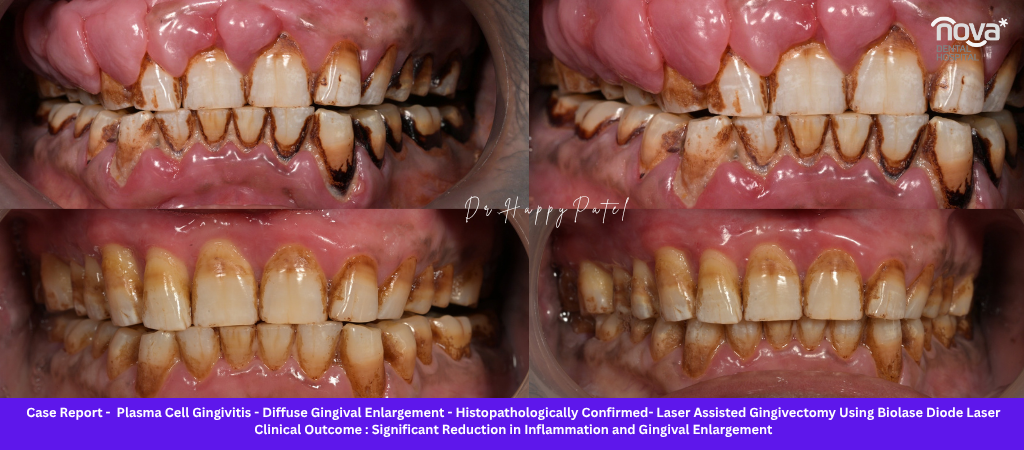
The patient who presented to our clinic demonstrated generalised redness, enlargement, and intense inflammation of the gum tissue across both the upper and lower jaws, accompanied by spontaneous bleeding and significant discomfort during routine brushing. The gum tissue was markedly swollen, erythematous, and friable — bleeding readily on contact with the periodontal probe during clinical examination.
A detailed dental and medical history revealed a long-standing habit of pan chewing extending over several years. Pan (betel leaf preparation) is well recognised as a source of chronic oral mucosal irritation — the chemical and mechanical irritation it produces in the oral soft tissues can sustain and amplify inflammatory responses, and its use has been associated with a range of mucosal conditions. While pan chewing is not the primary aetiology of Plasma Cell Gingivitis in the classical sense, its role as a chronic local irritant was considered clinically relevant in this patient’s case and was incorporated into the overall assessment and treatment plan.
The clinical findings, combined with the habit history, prompted a tissue biopsy for histopathological examination — the definitive diagnostic step that confirmed the diagnosis.
What Causes Plasma Cell Gingivitis? Understanding the Aetiology
The exact cause of Plasma Cell Gingivitis is not fully established in the dental literature, and in a proportion of cases no definitive trigger is ever identified despite thorough investigation. However, the most consistently associated causative factors fall into the following categories:
Hypersensitivity and Allergic Reactions
The most widely accepted aetiology for Plasma Cell Gingivitis is a Type IV (delayed-type) hypersensitivity reaction to an allergen in contact with the oral mucosa. Identified allergens include:
- Toothpaste ingredients — particularly flavouring agents such as cinnamon, spearmint, and peppermint, as well as preservatives and detergents (sodium lauryl sulphate)
- Mouthwash components — antiseptic agents and alcohol-based formulations
- Chewing gum additives — flavourings, sweeteners, and preservatives
- Herbal dental products — neem-based pastes, clove oil preparations, and other plant-derived oral care products used widely in India
- Food allergens — certain spices, particularly cinnamon and cardamom, have been implicated in documented cases
- Flavouring agents in confectionery and breath fresheners
The allergic hypothesis is supported by cases in which the condition resolves partially or completely following identification and elimination of the suspected allergen — the allergen patch testing equivalent of a clinical cure.
Chronic Local Irritation
Persistent mechanical or chemical irritation from local sources can sustain a chronic inflammatory infiltrate in the gingival connective tissue. Recognised local irritants include:
- Dental plaque and calculus — the bacterial biofilm and its mineralised derivative maintain a persistent inflammatory challenge at the gum margin
- Tobacco-related habits — smoking and smokeless tobacco products produce direct mucosal irritation
- Pan chewing — as seen in our case, the chronic irritation from betel leaf preparations and their additives (areca nut, slaked lime, tobacco) produces sustained mucosal trauma and chemical irritation
- Ill-fitting dentures or dental appliances that create chronic pressure or friction on the gum tissue
Idiopathic Cases
In a meaningful proportion of patients with histologically confirmed Plasma Cell Gingivitis, no specific allergen or irritant can be identified despite thorough investigation including patch testing and elimination protocols. These cases are classified as idiopathic and present a particular clinical challenge, as removing the causative trigger — the most clinically effective intervention — is not possible.
Why Accurate Diagnosis Is Critical — The Differential Diagnosis Challenge
Plasma Cell Gingivitis cannot be confidently diagnosed from its clinical appearance alone. This is the single most important clinical fact about the condition. The gum enlargement, redness, and bleeding it produces are features shared by a range of conditions — some of which are far more serious than Plasma Cell Gingivitis itself and require entirely different management.
The differential diagnosis for generalised gingival enlargement with plasma cell infiltration includes:
| Condition | Key Distinguishing Features |
|---|---|
| Plasma Cell Gingivitis | Dense plasma cell infiltrate; non-neoplastic; inflammatory |
| Leukaemia-related gingival enlargement | Abnormal leukocytes on histology; systemic haematological abnormalities |
| Oral lichen planus | Wickham’s striae; characteristic histological band-like infiltrate |
| Mucous membrane pemphigoid | Subepithelial blistering; immunofluorescence positive |
| Pemphigus vulgaris | Intraepithelial blistering; acantholytic cells; immunofluorescence positive |
| Drug-induced gingival overgrowth | History of phenytoin, ciclosporin, or calcium channel blocker use; fibrotic tissue |
| Chronic inflammatory gingival enlargement | Plaque-associated; responds to scaling; no dense plasma cell infiltrate |
The consequences of misdiagnosis in this differential are significant. Missing a leukaemic infiltrate means missing a systemic haematological malignancy. Confusing Plasma Cell Gingivitis with pemphigus vulgaris means initiating the wrong immunosuppressive regimen. Treating the condition as simple plaque-associated gingivitis means applying a treatment (scaling alone) that will not resolve the underlying plasma cell infiltrate.
Biopsy is therefore not a supplementary investigation — it is the definitive step that resolves this clinical uncertainty and directs the treatment appropriately.
Histopathological Findings — What the Biopsy Reveals
The tissue obtained at biopsy is sent for histopathological processing — preparation of thin tissue sections that are stained and examined under the microscope by a pathologist or oral pathologist.
In confirmed Plasma Cell Gingivitis, the microscopic examination reveals:
- Dense infiltration of mature plasma cells within the lamina propria (the connective tissue layer of the gum) — this is the pathognomonic feature that gives the condition its name
- Chronic inflammatory changes in the surrounding connective tissue, including oedema and increased vascularity
- Absence of malignant cells — the plasma cells, while numerous, are mature and non-atypical, distinguishing the condition from plasma cell malignancies such as multiple myeloma
- Absence of epithelial atypia — ruling out precancerous or malignant mucosal changes
- Non-neoplastic, non-specific chronic inflammatory pattern that is consistent with a reactive rather than neoplastic process
In our patient’s case, the biopsy specimen sent from Nova Dental Hospital was examined and reported as showing a non-neoplastic inflammatory lesion with prominent plasma cell infiltration — findings consistent with and diagnostic of Plasma Cell Gingivitis, and ruling out the more serious conditions on the differential.
This histopathological confirmation was the pivotal step that allowed the clinical team to proceed with appropriate treatment planning with confidence.
Treatment of Plasma Cell Gingivitis — A Staged, Comprehensive Approach
The management of Plasma Cell Gingivitis is multifactorial — addressing the possible causative trigger, controlling the local inflammatory environment, and managing the tissue changes that have resulted from the chronic inflammation. The approach at Nova Dental Hospital follows a structured, phased protocol:
Phase 1 — Allergen Identification and Elimination
The first intervention is a systematic review of all products that contact the oral mucosa — toothpaste, mouthwash, chewing gum, breath fresheners, herbal dental products, and dietary items. Patients are advised to switch to unflavoured, preservative-free toothpaste (without cinnamon, peppermint, sodium lauryl sulphate, or strong flavouring agents) and to avoid all mouthwashes and flavoured products for a trial period. If there is a demonstrable improvement following elimination, the allergen is implicated and avoidance becomes a long-term management strategy.
Where the allergen cannot be identified clinically, formal patch testing with standard oral allergen series can be arranged.
Phase 2 — Professional Periodontal Cleaning
Thorough scaling and root planing — removal of all supragingival and subgingival plaque and calculus — reduces the local bacterial inflammatory burden and creates the clean tissue environment needed to assess the underlying plasma cell infiltrate without the confounding effect of plaque-associated inflammation. Professional cleaning also improves patient comfort and oral hygiene compliance.
Phase 3 — Anti-Inflammatory and Medical Management
Depending on the severity of the presentation and the response to allergen elimination and scaling, topical anti-inflammatory therapies may be considered — including topical corticosteroids applied to the affected gum tissue. In more severe or refractory cases, systemic anti-inflammatory agents may be recommended in coordination with the patient’s physician.
Phase 4 — Habit Counselling and Lifestyle Modification
Any identified local irritants or harmful habits are addressed directly. In our patient’s case, comprehensive counselling regarding the cessation of pan chewing was provided as a central component of the treatment plan. The chronic mucosal irritation from long-term pan use was identified as a significant contributing factor, and the patient was advised on cessation strategies, the oral health consequences of continued pan use, and the importance of habit elimination for long-term treatment success.
Phase 5 — Surgical Management With Advanced Diode Laser Technology
When significant gingival enlargement persists following conservative management — as is commonly the case when the tissue has undergone substantial fibrotic or inflammatory change — surgical removal of the excess tissue is necessary. In our patient, diode laser-assisted gingivectomy was performed to remove the enlarged, inflamed gingival tissue and restore the normal gum contour.
Diode Laser Treatment for Plasma Cell Gingivitis — The Nova Dental Advantage
The use of advanced Diode Laser technology for the surgical management of Plasma Cell Gingivitis represents a significant advancement over conventional scalpel-based gingivectomy. At Nova Dental Hospital, Gandhinagar, the diode laser was used to precisely excise the inflamed gingival tissue with a level of control and patient comfort that conventional surgical techniques cannot match.
How the Diode Laser Works in Gum Surgery
The diode laser emits a focused beam of light energy at a wavelength that is preferentially absorbed by haemoglobin and melanin in the soft tissue. As the laser tip contacts the gum tissue, it simultaneously cuts the tissue and seals the blood vessels — the coagulation effect occurs in the same instant as the excision. The result is a surgical field that is largely bloodless, giving the surgeon excellent visibility throughout the procedure and reducing the patient’s bleeding during and after surgery.
The laser also produces a superficial bactericidal effect at the tissue margins, reducing the microbial contamination of the surgical wound — an effect that conventional scalpels cannot provide.
Clinical Benefits for the Patient
During the procedure:
- Minimal bleeding due to simultaneous tissue cutting and vessel coagulation
- Excellent surgical precision — the laser tip can be guided with fine accuracy along the gum margin
- Better visibility throughout the procedure due to the haemostatic effect
- Reduced need for sutures in many cases
After the procedure:
- Significantly reduced post-operative discomfort compared to conventional gingivectomy
- Faster healing — the sealed wound margin heals with less secondary infection risk
- Reduced inflammatory response in the healing tissue
- Reduced bacterial contamination at the surgical site
- Better final gingival contour and aesthetics — the precision of the laser allows more refined contouring of the gum margin than a scalpel
For the patient’s overall experience:
- Less anxiety — the absence of visible bleeding and the reduced noise of laser surgery compared to conventional instrumentation reduces the procedural fear many patients experience
- Shorter recovery period — most patients return to normal eating and oral hygiene within a few days rather than the longer recovery associated with conventional surgery
- Improved long-term aesthetics — the gum contour achieved with laser excision is typically more natural and more consistent along the full arch
Clinical Outcome at Nova Dental Hospital
Following the complete treatment protocol — allergen elimination, professional scaling, habit counselling, and diode laser-assisted gingivectomy — our patient demonstrated remarkable clinical improvement across all assessed parameters:
- Significant reduction in gingival enlargement — the excess tissue was removed and the gum contour restored to a normal, healthy profile
- Resolution of intense redness and inflammation — the fiery erythema characteristic of the active condition substantially resolved as the inflammatory burden was reduced
- Improved gingival contour — the laser-assisted surgical contouring produced a well-defined, aesthetically natural gum line
- Better oral hygiene access — with the enlarged, painful tissue removed, the patient was able to resume thorough brushing and flossing without discomfort
- Improved smile aesthetics — the normalisation of the gum tissue significantly improved the patient’s overall smile appearance and confidence
- Enhanced oral comfort — the burning and sensitivity that had characterised the pre-treatment presentation resolved substantially following treatment
Clinical photographs taken before and after the procedure documented the transformation — a before-and-after comparison that demonstrated not only the clinical effectiveness of the treatment but also the precision of the diode laser in reshaping the gum tissue.
Long-term care instructions given to the patient included: complete cessation of pan chewing; meticulous oral hygiene with appropriate, unflavoured toothpaste; avoidance of previously identified or suspected allergens; and attendance at regular follow-up appointments to monitor for any recurrence of the plasma cell infiltrate.
A Note for Dental Students and Clinicians — Key Learning Points
Plasma Cell Gingivitis occupies an important position in the differential diagnosis of generalised gingival enlargement and represents one of the conditions that tests a clinician’s discipline in not accepting the most obvious diagnosis without appropriate investigation.
The clinical lessons from this case:
- Always maintain a broad differential diagnosis for generalised gingival enlargement — the clinical appearance alone is insufficient to distinguish between conditions with dramatically different prognoses and management approaches
- Biopsy is essential when the gingival condition does not respond as expected to scaling and oral hygiene improvement, or when the clinical presentation has features — particularly the dense erythema and tissue character of Plasma Cell Gingivitis — that are not typical of common plaque-associated disease
- Systemic conditions must be excluded — leukaemic infiltration of the gingiva, in particular, can mimic Plasma Cell Gingivitis and carries life-altering implications if missed
- A detailed habit history is a diagnostic tool — tobacco use, pan chewing, betel nut use, and unusual dietary or oral hygiene habits are all relevant to the aetiology and should be documented thoroughly
- Allergen identification requires systematic methodology — changing one product at a time, observing the response over a defined period, and maintaining records of the trial are the foundations of a meaningful allergen elimination protocol
- Diode laser surgery is an effective and patient-centred modality for the surgical phase of management, with documented advantages in haemostasis, precision, post-operative comfort, and healing outcomes
- Long-term follow-up is not optional — Plasma Cell Gingivitis can recur, particularly if the causative trigger has not been fully eliminated, and regular monitoring allows early identification of recurrence before the condition re-establishes at its previous severity
When Should You See a Dentist About Your Gums?
Plasma Cell Gingivitis is rare — but the gum symptoms it produces are shared by several conditions that all warrant professional evaluation. If you are experiencing any of the following, a dental assessment is appropriate without delay:
- Persistent gum swelling that has not improved with improved brushing and flossing
- Gums that appear intensely red — significantly more so than the mild pink-red of ordinary gum inflammation
- Repeated bleeding from the gums during or between brushing
- Gum enlargement that is beginning to cover the tooth surfaces
- A burning or stinging sensation in the gum tissue, particularly when eating
- Gum changes that have been present for several weeks without any obvious cause
Early diagnosis of any gum condition — whether it is common gingivitis or a rarer condition like Plasma Cell Gingivitis — allows the simplest, most effective treatment to be applied before the condition becomes more established. Delayed diagnosis means more complex treatment, less predictable outcomes, and a longer recovery.
Laser Gum Treatment in Gandhinagar — Nova Dental Hospital
At Nova Dental Hospital, Gandhinagar, the management of complex and rare gum conditions is supported by the full diagnostic and therapeutic infrastructure needed for accurate, comprehensive care:
- Detailed clinical assessment and history-taking — including thorough documentation of habits, medications, oral care products, and dietary factors relevant to the gum condition
- Biopsy and histopathological evaluation — for any gum condition that does not present or respond as expected, tissue biopsy is available and arranged at the clinic
- Advanced Diode Laser technology — for laser-assisted gingivectomy, gum recontouring, and management of gingival enlargement with the clinical advantages of precision, haemostasis, and patient comfort described above
- Comprehensive periodontal care — scaling, root planing, supportive periodontal therapy, and long-term maintenance for all stages of gum disease
- Patient education and habit counselling — addressing the behavioural and lifestyle factors that contribute to gum conditions, with specific guidance on allergen elimination, habit cessation, and oral hygiene optimisation
Whether the presentation is a straightforward case of gum inflammation or a complex, biopsy-confirmed rare condition, the clinical approach at Nova Dental Hospital is the same: accurate diagnosis first, then the most appropriate and patient-centred treatment available.
Frequently Asked Questions
Q: Is Plasma Cell Gingivitis contagious?
No. Plasma Cell Gingivitis is an inflammatory condition — most likely an immune reaction to an allergen or chronic irritant — and is not infectious or transmissible between individuals.
Q: Can Plasma Cell Gingivitis resolve on its own?
Rarely, if ever, without intervention. If the causative allergen is identified and eliminated very early, some improvement may occur. In established cases with significant gingival enlargement, professional treatment — including laser-assisted surgery in appropriate cases — is necessary for resolution.
Q: How long does recovery take after diode laser gum treatment?
Most patients notice significant improvement in comfort within two to three days of laser treatment. The gum tissue heals progressively over two to four weeks. The absence of sutures in many laser cases and the sealed wound margins make the recovery period considerably more comfortable than conventional surgical gingivectomy.
Q: Will Plasma Cell Gingivitis come back after treatment?
Recurrence is possible, particularly if the causative allergen or irritant has not been fully eliminated. This is why long-term follow-up and patient adherence to the habit and allergen avoidance advice are integral to the treatment plan — not supplementary recommendations.
Q: Does diode laser treatment hurt?
The procedure is performed under local anaesthesia and should not be painful during treatment. Post-operative discomfort is typically mild and well-managed with standard pain relief. The laser’s coagulation effect means the wound margins are sealed, which significantly reduces post-operative sensitivity compared to conventional surgery.
Dr. Happy Patel (BDS) Nova Dental Hospital, Gandhinagar Advanced Laser Dentistry | Painless Dental Care | Comprehensive Gum Treatment
For appointments: +91 9638 111 082 | Mon–Sat: 9 AM–9 PM | Sun: 9:30 AM–1:30 PM Nova Dental Hospital — Laser Gum Treatment, Gandhinagar
Related posts

Write a Comment
Recent Posts


