
Radiation Safety: The Truth About 3D Dental Scans
Radiation Safety: The Truth About 3D Dental Scans
The Question Every Patient Deserves an Honest Answer To
“Is this scan safe?”
It is one of the most common questions patients ask before a 3D dental scan — and it is exactly the right question to ask. Radiation is invisible, and when a machine rotates around your head, it is completely natural to feel uncertain about what is actually happening.
The honest answer is this: a CBCT dental scan is safe. But that answer means more when you understand why — when you can see the actual numbers, compare them to things you already experience, and understand the precautions that responsible dental clinics put in place for every single scan.
This blog is written to give you that understanding. Not to dismiss your concerns, and not to replace the conversation you should have with your dentist — but to give you the factual foundation to ask better questions and feel genuinely confident when advanced dental imaging is part of your treatment plan.
🔑 Key Takeaways
- CBCT dental scan safety is well-established — doses are a fraction of what patients often fear.
- A CBCT scan typically delivers less radiation than a day of natural background exposure at altitude.
- The ALARA principle — As Low As Reasonably Achievable — governs every responsible dental imaging decision.
- Radiation dose varies significantly by field of view; smaller scans mean meaningfully lower exposure.
- Special considerations apply for children and pregnant patients — and good dentists always account for them.
- The risk of an undiagnosed dental condition far outweighs the minimal risk of a clinically justified CBCT scan.
What Is Dental Radiation — and Why Does It Matter?
Radiation is energy moving through space. It surrounds us every day — from the sun, the ground beneath our feet, the food we eat, and even the walls of the buildings we live in. This constant, unavoidable exposure is called natural background radiation, and for most people it amounts to approximately 2,000 to 3,000 microsieverts (μSv) per year.
When dentists and radiologists talk about radiation safety, the unit they use is the microsievert (μSv) — a measure of the biological effect of radiation on human tissue. The lower the μSv, the lower the biological impact. Context matters enormously here, because many patients imagine dental X-ray doses far higher than they actually are.
Understanding where dental radiation sits within the broader picture of everyday exposure is one of the most reassuring things you can do before any scan — so that is exactly where we will start.
Putting Dental Radiation in Context
The table below places common dental imaging doses alongside everyday radiation sources to give you a realistic sense of proportion:
| Source of Radiation | Approximate Dose (μSv) | Equivalent To |
| Eating 10 bananas | 0.1 μSv | Natural potassium-40 in food |
| Single dental bitewing X-ray | 1–8 μSv | 1–4 hours of background radiation |
| OPG (full jaw panoramic X-ray) | 14–24 μSv | 1–3 days of background radiation |
| Chest X-ray | 20 μSv | ~2–3 days of background radiation |
| CBCT — small field of view | 40–100 μSv | ~4–10 days of background radiation |
| CBCT — large field of view | 200–600 μSv | ~3–8 weeks of background radiation |
| Transatlantic flight (London–NY) | 50–80 μSv | Cosmic radiation at altitude |
| Annual background radiation (average) | 2,000–3,000 μSv | Baseline for everyone, every year |
| Chest CT scan (medical) | 5,000–7,000 μSv | ~2–3 years of background radiation |
| Annual occupational limit (radiation workers) | 20,000 μSv | Regulatory maximum for professionals |
Even a large-field CBCT scan — the highest-dose scenario in routine dental imaging — represents less than one month of the natural background radiation every person absorbs simply by living on Earth.
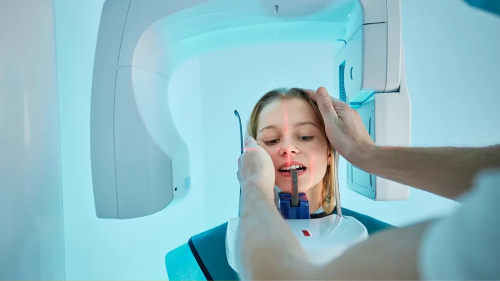
How a CBCT Scan Actually Delivers Radiation
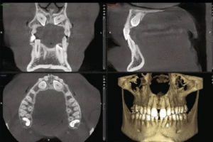
A CBCT dental scan works by rotating a cone-shaped X-ray beam 360° around the patient’s head, capturing hundreds of individual projection images in a single sweep. These images are reconstructed by software into a three-dimensional volume — the precise anatomical map that makes CBCT so valuable for implant planning, complex extractions, and advanced root canal treatment.
The radiation is delivered only during this single rotation, which typically takes between 10 and 40 seconds. There is no residual radiation after the scan is complete, no radioactive material introduced into the body, and no lasting effect on the tissues exposed.
What determines the dose a patient receives is primarily driven by three factors:
Field of View (FOV)
The field of view is the size of the anatomical region being scanned. A small FOV targeting a single tooth or implant site delivers far less radiation than a large FOV capturing the entire skull. This is one of the most important dose-reduction levers available to your dental team — and one that responsible clinicians always use.
At Nova Dental Hospital, the field of view is always selected based on what the clinical case actually requires — never defaulting to a larger scan than necessary.
Exposure Parameters
Tube voltage (kV) and tube current (mA) are the technical settings that control the intensity of the X-ray beam. Modern CBCT units allow these to be adjusted to the minimum level needed to produce a diagnostically adequate image for the specific patient and the specific task. Smaller patients and paediatric cases always warrant reduced parameters.
Number of Projections and Rotation Arc
Some CBCT protocols use a full 360° rotation while others use a partial arc of 180° or less. Reduced-arc protocols can meaningfully lower dose while still generating sufficient diagnostic quality for many clinical tasks. The clinical indication determines which protocol is appropriate.
✅ Quick Tip: Questions to Ask Before Your CBCT Scan
- “What field of view are you using, and is it the smallest appropriate for my case?”
- “Has the exposure been adjusted for my size and age?”
- “Is a CBCT necessary for this procedure, or would an OPG give you sufficient information?”
- “Can I see the images and have them explained to me after the scan?”
The ALARA Principle — The Standard Every Dental Clinic Should Follow
ALARA stands for As Low As Reasonably Achievable — and it is the governing principle of radiation protection in medicine and dentistry worldwide. It is not simply a guideline; it is the ethical and clinical standard by which every diagnostic imaging decision should be evaluated.
ALARA means that before any scan is ordered, the clinician must weigh two things against each other: the diagnostic benefit of the information the scan will provide, and the radiation dose the patient will receive to obtain it. A scan is only justified when the benefit clearly outweighs the dose.
In practice, this means:
- No routine CBCT scans — a 3D dental scan should never be ordered as a standard yearly check-up tool. Each prescription requires a specific clinical justification.
- Smallest appropriate FOV — if only the lower left molar region needs to be assessed, the scan should not include the entire skull.
- Patient-specific parameters — dose settings should be adjusted for the individual patient’s size, age, and the anatomical region being imaged.
- Alternative imaging considered first — in many cases, an OPG or periapical X-ray can answer the clinical question at a fraction of the dose. CBCT is reserved for when 3D information is genuinely necessary.
At Nova Dental Hospital’s imaging facility, ALARA is embedded in every imaging decision. Our radiologist and dental specialists review the clinical indication before any scan is prescribed — and when a simpler imaging modality can answer the clinical question, it is chosen.
CBCT Dental Scan Safety: How It Compares to Other Medical Imaging
One reason patients sometimes overestimate the risk of a CBCT is that it shares a name with the CT scanner used in hospitals — and medical CT scans do carry higher radiation doses. The distinction is important.
A hospital CT scan of the chest or abdomen uses a fan-shaped beam designed to image large body regions and typically delivers 5,000 to 10,000 μSv — sometimes significantly more. A dental CBCT uses a narrowly focused cone beam targeting only the jaw and teeth, operating at a fundamentally lower dose range.
The comparison below illustrates how CBCT dental scan safety stacks up against other common medical imaging procedures:
| Imaging Procedure | Typical Dose Range | Clinical Context |
| Dental periapical X-ray | 1–8 μSv | Single tooth, very low dose |
| OPG (full jaw panoramic) | 14–24 μSv | Full arch overview, routine screening |
| CBCT small FOV (1–2 teeth) | 40–100 μSv | Implant site, specific RCT planning |
| CBCT medium FOV (one jaw) | 100–200 μSv | Surgical planning, multiple implants |
| CBCT large FOV (full skull) | 200–600 μSv | Orthognathic, TMJ, complex cases |
| Chest X-ray | 20 μSv | Standard medical imaging |
| Lumbar spine X-ray | 700 μSv | Orthopaedic assessment |
| Medical CT head | 2,000 μSv | Neurological or trauma assessment |
| Medical CT chest/abdomen | 5,000–10,000 μSv | Oncology, trauma, organ assessment |
| Annual background radiation | 2,000–3,000 μSv | Unavoidable baseline for all people |
A small-field CBCT scan — the type used for most single-implant planning — delivers a dose comparable to a short-haul flight, or roughly one week of normal background radiation. This context is what matters when evaluating CBCT dental scan safety in real clinical terms.
Special Considerations: Children, Pregnancy, and Frequent Imaging
Children and Paediatric Patients
Children are more sensitive to radiation than adults — their cells divide more rapidly, and they have more years ahead in which any theoretical cumulative effect could manifest. This does not mean CBCT is unsafe in children; it means that clinical justification must be even more rigorous, and dose reduction measures even more carefully applied.
For paediatric patients, responsible dental imaging practice includes:
- Using the smallest possible field of view that answers the clinical question
- Reducing tube current and voltage relative to adult settings
- Avoiding full-skull large-FOV scans unless absolutely required for complex surgical planning
- Considering whether an OPG or periapical X-ray can provide equivalent diagnostic information at lower dose
- Ensuring the child is able to remain still during the scan to avoid repeat exposures
At Nova Dental Hospital, our paediatric dental team follows age-specific imaging protocols — and CBCT is only prescribed for children when it is clinically unavoidable and clearly in the child’s best diagnostic interest.
Pregnancy
Dental X-rays during pregnancy are generally considered safe at the doses used in routine practice — including OPG. The developing foetus is located far from the field of dental X-ray exposure, and the use of a lead apron (which is standard practice) further reduces scatter radiation to effectively negligible levels.
That said, the approach at Nova Dental Hospital is to avoid CBCT scans during the first trimester wherever possible, as this is the most sensitive period of foetal development. During the second and third trimesters, clinically necessary imaging can be performed with lead apron protection and careful dose minimisation.
If you are pregnant or planning to become pregnant, always inform your dentist before any imaging appointment. Non-urgent dental work and imaging is almost always deferrable, and we will always discuss your options transparently.
Patients Who Require Repeated Imaging
Some patients — particularly those undergoing full-mouth implant rehabilitation or complex orthodontic treatment — may require more than one CBCT scan across their treatment timeline. Even in these cases, cumulative doses remain well within internationally recognised safety thresholds. Each additional scan is only prescribed when the clinical information it provides genuinely changes or advances the treatment plan.
The important point is that no imaging is ever prescribed out of habit or routine. Every scan has a clinical question it is designed to answer.
✅ Quick Tip: Reducing Your Own Exposure Sensibly
- Always ask your dentist whether the planned imaging is necessary for your specific treatment — it is a reasonable and welcome question.
- If you have had recent dental X-rays at another clinic, bring those images or request their transfer before having new scans taken.
- Do not decline necessary imaging out of radiation concern alone — the diagnostic benefit of finding an infection, locating a nerve, or measuring bone accurately almost always outweighs the minimal dose.
- Wear a lead apron when offered — it is standard practice and adds a meaningful layer of protection.
Common Misconceptions About 3D Dental Scan Radiation
This is one of the most common and understandable misconceptions. The word ‘CT’ appears in both names, but the technologies and their doses are fundamentally different. A medical CT scan of the chest or abdomen can deliver 50 to 250 times more radiation than a small-field dental CBCT. The cone beam technology in dental CBCT was specifically developed to capture the precision of 3D imaging at a fraction of the dose required by conventional medical CT.
Radiation exposure does accumulate, but the relevant comparison is always against natural background radiation — which also accumulates continuously throughout life. The additional lifetime dose from a typical course of dental imaging, including occasional CBCT scans, is negligibly small relative to the 2,000–3,000 μSv per year that every person receives from nature. The body’s repair mechanisms also handle low-level radiation exposure effectively.
This position, while understandable, carries its own risk. Undiagnosed dental infections, missed root canal anatomy, an implant placed without 3D bone mapping, or a cyst that goes undetected — these are real clinical harms. The purpose of diagnostic imaging is to prevent treatment errors and catch problems early. Refusing clinically indicated imaging does not eliminate risk; it simply shifts the risk from a theoretical radiation concern to a concrete diagnostic one.
This is incorrect in both directions. Higher radiation does not automatically improve image quality — modern CBCT systems are engineered to produce diagnostically excellent images at the lowest achievable dose. Increasing dose beyond what is clinically necessary does not enhance the scan; it only increases exposure without benefit. This is precisely why ALARA principles and dose optimisation are so important.
Dental X-rays are routinely and safely used in children across the world. The British Society of Dental and Maxillofacial Radiology, the American Academy of Pediatric Dentistry, and the European Commission all provide paediatric-specific guidelines that support clinically justified imaging with appropriate dose reduction. Undetected decay, infection, or developmental abnormalities in children carry far greater long-term consequences than the carefully managed doses used in modern dental radiography.
How Nova Dental Hospital Approaches Radiation Safety
Radiation safety at Nova Dental Hospital is not a box-ticking exercise — it is a clinical philosophy built into every imaging decision. Our in-house radiology suite was designed with both diagnostic quality and patient safety as equal priorities.
Our imaging protocols reflect the following commitments:
- Clinician-prescribed imaging only — no CBCT or OPG is ordered without a specific clinical indication documented by the treating dentist.
- FOV selection per case — the field of view is chosen to cover only the anatomical region relevant to the clinical question, never larger.
- Dose parameters adjusted per patient — children, smaller adults, and patients with specific sensitivities receive individually calibrated exposure settings.
- Lead apron protection as standard — protective shielding is provided for every patient, every time.
- Immediate specialist review — because our imaging is in-house, results are reviewed by the treating specialist in the same appointment, eliminating the need for repeat scans due to communication gaps.
- Transparent communication — patients are always told which type of scan is being taken, why it is necessary, and what the approximate dose involves.
You can read more about the full range of imaging technology available at our facility — including how OPG and CBCT serve different clinical purposes — on our CBCT and OPG imaging page.
Patients who have visited us and want to share their experience are also welcome to leave a review on our Google Business Profile — it genuinely helps other patients in Gandhinagar make informed decisions about their dental care.
When Is a CBCT Dental Scan Genuinely Justified?
Given that CBCT carries a higher dose than a standard OPG, the decision to prescribe it is made carefully. The following table summarises the clinical scenarios where CBCT provides diagnostic value that justifies its use, and those where simpler imaging is sufficient:
| Clinical Scenario | Appropriate Imaging | Reason |
| Routine annual dental check-up | OPG or periapical X-rays | Overview imaging at minimal dose is sufficient |
| Single tooth extraction (erupted) | Periapical X-ray | Local 2D view adequate |
| Dental implant planning | CBCT (small/medium FOV) | Bone volume, nerve distance, and density required in 3D |
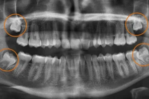
| Wisdom tooth removal (near nerve) | CBCT | 3D nerve proximity essential to prevent injury |
| Wisdom tooth removal (straightforward) | OPG | 2D panoramic view sufficient |
| Root canal — single root, first treatment | Periapical X-ray | Standard 2D adequate for uncomplicated cases |
| Root canal — complex, retreatment | CBCT (small FOV) | Canal morphology, periapical lesion extent in 3D |
| Orthodontic planning (impacted teeth) | OPG + CBCT (targeted) | OPG for overview; CBCT to localise impaction in 3D |
| Orthodontic planning (standard) | OPG | Full arch development assessment in 2D sufficient |
| TMJ pain assessment | OPG first; CBCT if bone changes suspected | Escalate to 3D only when initial imaging indicates need |
| Jaw cyst or tumour evaluation | CBCT | Boundary mapping and internal structure in 3D required |
| Paediatric growth screening | OPG (dose-reduced) | Full arch eruption overview; avoid CBCT unless essential |
Frequently Asked Questions
FAQ 1: Is a CBCT dental scan safe for regular use?
CBCT is not designed for regular or routine use — it is a diagnostic tool prescribed when 3D information is clinically necessary. When used appropriately, it is safe. The key safeguard is clinical justification: a CBCT should only be ordered when it will provide information that an OPG or conventional X-ray cannot, and when that information is essential for treatment planning. Used this way, the diagnostic benefit is clear and the radiation risk is minimal.
FAQ 2: How does CBCT radiation compare to a flight?
A short-haul flight typically exposes a passenger to approximately 20–50 μSv of cosmic radiation from altitude. A small-field CBCT scan used for a single implant site delivers roughly 40–100 μSv — comparable to one or two short flights. A transatlantic flight delivers approximately 50–80 μSv. These comparisons are not meant to trivialise radiation exposure, but to provide genuine context: the doses involved in dental CBCT imaging are within the range of everyday activities that people undertake without concern.
FAQ 3: Should I be worried if my dentist recommends a 3D dental scan?
No — and in fact, if your dentist is recommending a CBCT, it is because they need precise 3D information to treat you safely and accurately. The scenarios where CBCT is most commonly used — implant placement, complex extractions near nerves, retreatment of difficult root canals — are precisely the cases where the cost of insufficient diagnostic information is highest. A dentist who recommends a CBCT when clinically justified is being thorough, not reckless.
FAQ 4: Can I ask my dentist to use a lower-dose protocol?
Yes — and it is a reasonable thing to ask. A good dental practice will already be using the lowest dose protocol appropriate for your specific clinical case. You can ask which field of view is being used and whether a smaller FOV is possible. You can ask about the exposure parameters and whether they have been adjusted for your size. At Nova Dental Hospital, dose optimisation is standard practice — but we always welcome the opportunity to walk patients through the imaging decision and explain the rationale.
FAQ 5: Does Nova Dental Hospital follow international radiation safety guidelines?
Yes. Our imaging protocols are aligned with the guidelines set by the European Commission’s cone beam CT radiation protection criteria, the recommendations of the British Society of Dental and Maxillofacial Radiology, and the International Atomic Energy Agency’s dental imaging safety standards. The ALARA principle governs every scan prescription. Our in-house radiologist reviews clinical indications before imaging is approved, and our CBCT system is maintained and calibrated regularly to ensure dose accuracy and image quality.
🔑 Key Takeaways
- CBCT dental scan safety is supported by decades of clinical evidence — doses are low and well within internationally accepted thresholds.
- A small-field CBCT scan delivers roughly the same radiation as a short-haul flight or a week of natural background exposure.
- The ALARA principle requires that every scan be clinically justified, using the smallest appropriate field of view and optimised parameters.
- Children and pregnant patients warrant additional caution, but are not categorically excluded from dental imaging when it is genuinely necessary.
- Refusing clinically indicated imaging is not a risk-free choice — missed diagnoses carry their own consequences.
- Nova Dental Hospital prescribes CBCT only when 3D information is essential, using in-house specialist review to make imaging decisions accurately and immediately.
Informed Consent Begins With Informed Patients
Radiation safety in dentistry is a genuinely important topic — not because dental X-rays are dangerous, but because patients deserve to understand what is happening and why. Fear that is rooted in misunderstanding leads to avoidance, and avoidance of necessary diagnostic imaging leads to missed diagnoses, delayed treatment, and worse outcomes.
The truth about CBCT dental scan safety is this: when prescribed for the right clinical reason, using the smallest appropriate field of view, with dose parameters optimised for the individual patient, a 3D dental scan is one of the most valuable and clinically safe diagnostic tools in modern dentistry. It allows your dental team to treat you with a level of precision, foresight, and safety that was simply not possible a generation ago.
At Nova Dental Hospital in Gandhinagar, every imaging decision is made with your safety and your clinical outcomes as the guiding priorities. If you have questions about a scan that has been recommended for you, we encourage you to ask them — before, during, and after your appointment. You can also explore our full CBCT and OPG imaging facility page to understand the technology and protocols we use.
Related posts
Write a Comment
Recent Posts

Radiation Safety: The Truth About 3D Dental Scans

CBCT vs. OPG: Understanding Full Jaw 3D Imaging

3D Scan for Root Canal: Finding Hidden Canals

